Hello steemit community, I am a graduate student in microbiology, I am currently working in a clinical laboratory, which is qualified to study and learn in areas, including hematology, parasitology, bacteriology and biochemistry, I want to share some of the microorganisms that analyze in the laboratory.
Today I will show you the parasitology area part 1:
Exam: croproanalysis
Analysis: microscopic
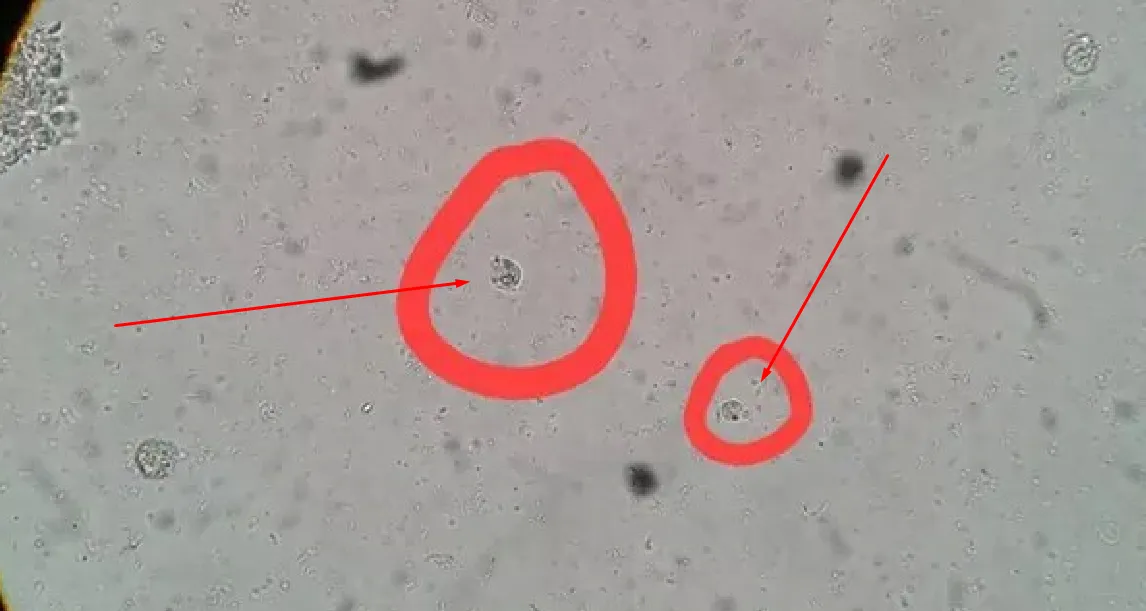
1.-Entamoeba coli:
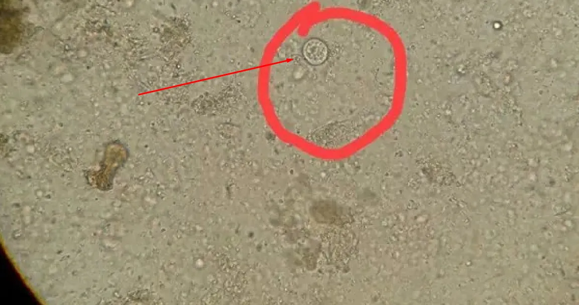
2.-Entamoeba histolitica/dispar:
3.-Balantidium coli
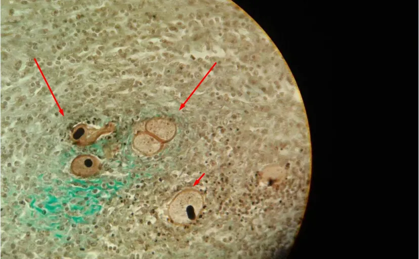
Observations:
-The name of the parasite must be written in italics as established by the binominal nomenclature of microorganisms and parasites
-The photos were taken from micrographs taken at 100X with a Zeiss photomicroscope, model FL 40. The analysis of the micrographs taken from the plates was made with the program Adobe Photo Deluxe.
I hope your help, these parasites are very common, many times they are asymptomatic, so I recommend taking the pictures that are very simple.
Until the next delivery.