Hello everyone, how are you all? I hope you all are doing well. I am too. As i am posted in microbiology department for 15 days they have given me schedule and taking classes. Yesterday class was taken on fungi by mam and she asked me to see a slide.
Case
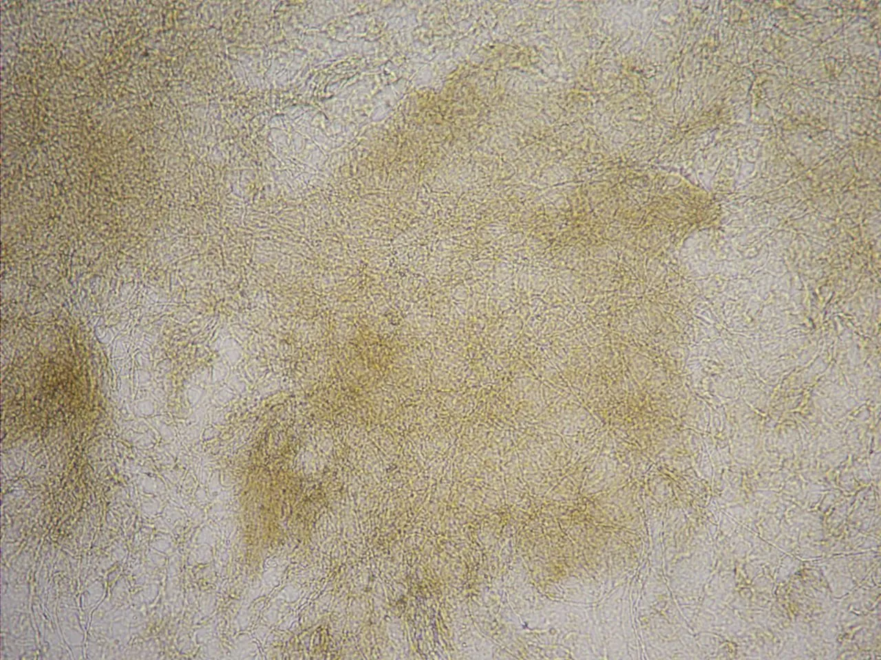
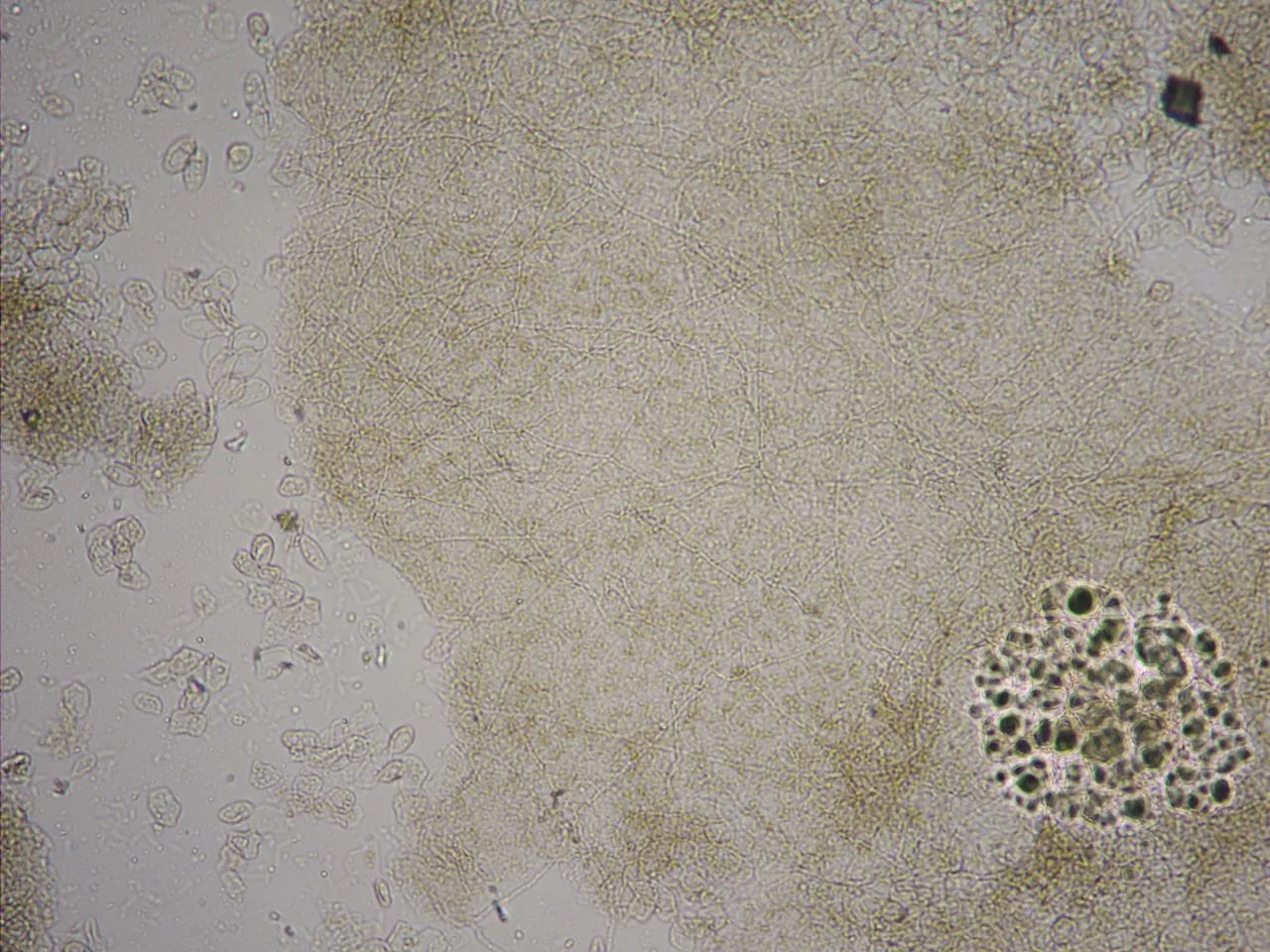
Patient came with complaints of lesions over the palms. Sample was taken from the lesion and KOH mounting was done. This is what i have seen on microscopy -
This was examined under 10X magnification only. As the microscope is connected to the computer system with procam. I am abe to take rectangular picture.
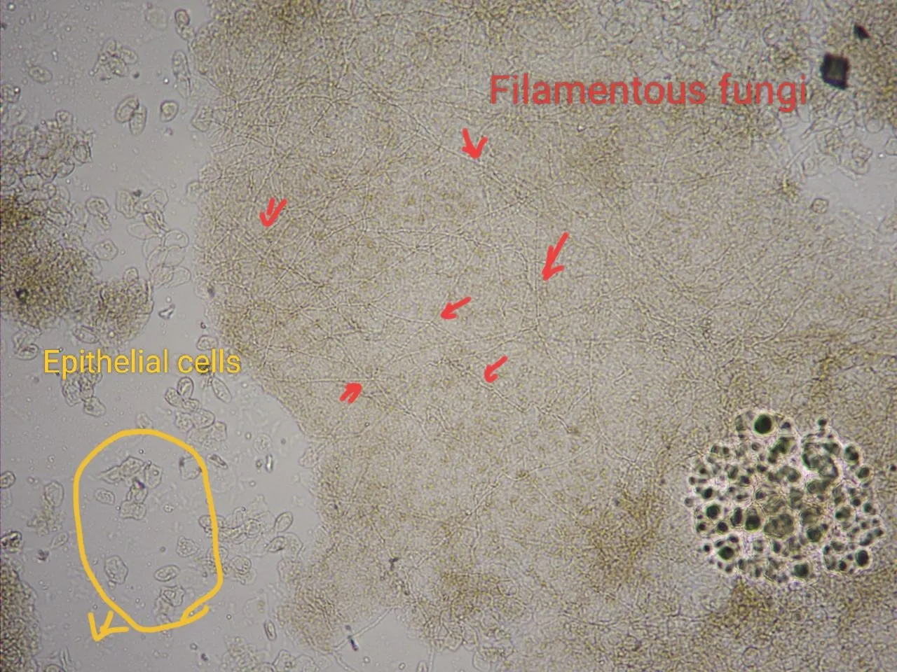
I have labelled the filamentous fungi with red arrows. See the filamentous fungi and tgere are epithelial cells which i labelled with yellow color.
So we are suspecting it as Tinea manuum why because of this microscopic features and the site of lesion which is on palms.
So this patient will be suggested to go for antifungal drugs.
References
- Ananthanarayan and Paniker's Textbook of Microbiology, Eleventh Edition
Thanks for reading,
with regards,