Our digestive system is a marvel of biological engineering, and understanding its intricacies can shed light on how our bodies process the food we consume. In this post, we'll embark on a journey through the digestive system, exploring its various organs, functions, and processes.
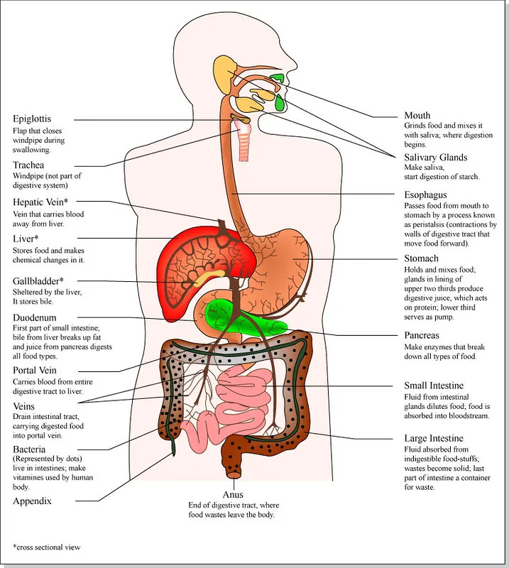
Our digestive journey commences in the oral cavity, where the process of digestion begins. Teeth play a crucial role in mastication, breaking down food into smaller pieces and this is a vital step in physical digestion. As we chew, saliva moistens the food and transforms it into a soft mass known as bolus. This is where amylase in saliva initiates the digestion of carbohydrates. Within the oral cavity, several glands, including the parotid, sublingual, and submandibular glands, produce saliva. Of these, the submandibular gland is the most prolific in generating saliva, followed by the parotid and sublingual glands.
Flickr
After the saliva has mixed the food, the food is transferred to the esophagus which transport food from the oral cavity to the stomach. In this process, nothing happens to the food being swallowed. The esophagus is made up of three muscle types which are the skeletal muscle, the mixed muscle, and the smooth muscle. Proximally, the skeletal muscle lines the esophagus from the pharynx to the top of the esophagus. It allows us to voluntarily swallow food.
The proximal distal part of the esophagus is made up of mixed muscle which is made up of both the skeletal and smooth muscle. At the distal part of the esophagus that goes to the stomach is made of smooth muscle which functions involuntarily. This means that when we swallow food, we go from voluntarily to involuntarily movement which is known as peristalsis.
The gastrointestinal (GI) tract consists of four layers: mucosa, submucosa, muscularis externa, and serosa. The mucosa lines the inner lumen and serves roles in protection, absorption, and secretion. Simple columnar epithelium and the lamina propria are components of the mucosa, with the latter housing systemic capillaries for nutrient absorption.

Below the mucosa is the submucosa, which contains numerous vessels that nourish the mucosa. The muscularis mucosa, a thin layer of smooth muscle, separates the mucosa from the submucosa. The muscularis externa, consisting of inner circular and outer longitudinal smooth muscle layers, facilitates peristalsis and segmentation of food. Nerve plexuses, like the Auerbach's and Meissner's plexuses, coordinate GI tract functions. Lastly, the serosa is a mesothelial lining that produces serous fluids.
The stomach is the next location where the food arrives after leaving the esophagus. The stomach is located between the esophagus and the duodenum. It is superficial to the abdominal aorta, the spleen, and the pancreas. In the stomach, peristalsis causes the bolus to become chyme which then helps in the mixing of the food.
In the stomach, Goblet Cells make mucous which prevents the stomach from the acid being produced in the stomach by the parietal cells which produces hydrochloric acid and Intrinsic factor. The intrinsic factor helps with the reabosoption of vitamin B12 in the small intestine. The chief calls produces pepsinogen which becomes pepsin when mixed with hydrochloric acid for the digestion of protein.
The stomach's anatomical features include the cardiac sphincter, cardia, fundus, body, and pylorus, which houses the pyloric sphincter, responsible for regulating the passage of contents to the small intestine.
Next on our journey is the small intestine, roughly 7 meters in length. Despite its name, the "small" intestine refers to its narrow lumen diameter, not its length. It consists of three segments: the duodenum, jejunum, and ileum. Here, digestion is completed, and nutrient absorption occurs.
The Duodenum is the first part of the small intestine that has the bruner's gland and has four parts which as the superior part (made up of the duodenal cap/bulb which has an intraperitoneal smooth wall), Other parts of the duodenum are retroperitoneal and they include the second part of the duodenum which is the descending part of the duodenum which has circular fold, and has the major duodenal papilla. At the part, common bile duct and the common pancreatic duct form which helps for bile and pancreatic enzyme to get to the duodenum.
The third part of the duodenum is the transverse part of the duodenum where the superior mesenteric artery and superior mesenteric vein crosses the duodenum. The ascending part of the duodenum is the fourth part of the duodenum, and it is where the duodenal regional junction occurs, and it is has an intraperitoneal smooth wall. In the fourth part is where the ligament of Treitz comes to form the duodenojejunal junction.
Moving along, the jejunum and ileum are responsible for nutrient absorption. The jejunum, located in the upper left quadrant, boasts numerous circular folds (plicae circulares) for efficient absorption. In contrast, the ileum, found in the lower right quadrant, features fewer circular folds.
Our journey concludes in the large intestine, which is about 1.5 meters long and characterized by its large lumen diameter. Notable features include Taeniae coli, haustra, and epiploic appendages. The large intestine plays a crucial role in absorbing water, salts, and vitamins produced by intestinal bacterial flora. It also compacts and eliminates feces.
The Cecum is located at the lower right quadrant of the abdomen and is a blind-ended sac, and its starts the large intestine. The vermiform appendix is attached to the cecum. The next part of the intestine is the Ascending colon which arises from the cecum and goes vertically to the liver, towards the right side of the abdomen. The transverse colon is the superior part of the abdomen and an intraperitoneal organ courses from the right colic flexor to the left colic flexor and it is anchored to the posterior abdominal wall by the transverse muscle colon. The decending colon goes from the left colic or splenic flexure to the sigmoid colon.
The rectum is the straight part of the gastrointestinal tract. The anus comes after the rectum, and the anus is the exit of the gastrointestinal tract. It is made up of a skeletal muscle which makes up the external anal sphincter which helps to control the exist of waste from the body.
The digestive system is a marvel of well structured biological engineering, orchestrating a series of complex processes to break down and absorb nutrients essential for our survival. So, as you savor your next meal, remember the incredible journey it embarks on within your body. In the next post, I will be discussing the accessory digestive organs and their contribution to digestion, and the vascular supply and innervation of the GI tract.
Reference
https://training.seer.cancer.gov/anatomy/digestive/structure.html
https://www.ncbi.nlm.nih.gov/books/NBK54098/
https://www.ncbi.nlm.nih.gov/books/NBK542251/
https://www.kenhub.com/en/library/anatomy/the-duodenum
https://www.ncbi.nlm.nih.gov/pmc/articles/PMC6825871/
https://www.ncbi.nlm.nih.gov/books/NBK544242/
https://www.ncbi.nlm.nih.gov/pmc/articles/PMC4956471/
https://www.ncbi.nlm.nih.gov/books/NBK470577/
https://www.ncbi.nlm.nih.gov/books/NBK482390/
https://www.msdmanuals.com/home/digestive-disorders/biology-of-the-digestive-system/rectum-and-anus