Greetings, friends of Hive
For March, the Med-Hive community has chosen as a theme Women's health, I invite you to join the participation, here is the link.
When we refer to the female anatomy, we mean the whole body. However, medically we say "female organs" to the external and internal gynecological organs. These structures are important because they are involved in fertility, reproduction, menstruation, and sexual activity.
Female Internal Genitals
The female reproductive system (internal genital organs) consists of the ovaries, fallopian tubes, uterus, cervix, and vagina.
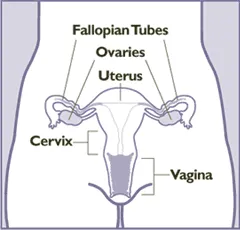
Female internal genitalia. Frontal view.
Credit: Centers for Disease Control and Prevention Public domain.
Ovaries
These are the sex glands. They are oval and approximately 3 cm in size. They are located inside the pelvis, on either side of the uterus.
Their two main functions are to produce hormones (estrogen and progesterone) and to release eggs.
The ovaries contain within them follicles and within the follicles are the immature eggs. Each month, a follicle matures, ruptures, and releases an egg (female sex cell), this process is called ovulation. The mature egg will go to the uterus and implant for possible fertilization. If fertilization does not occur, the egg will be destroyed.
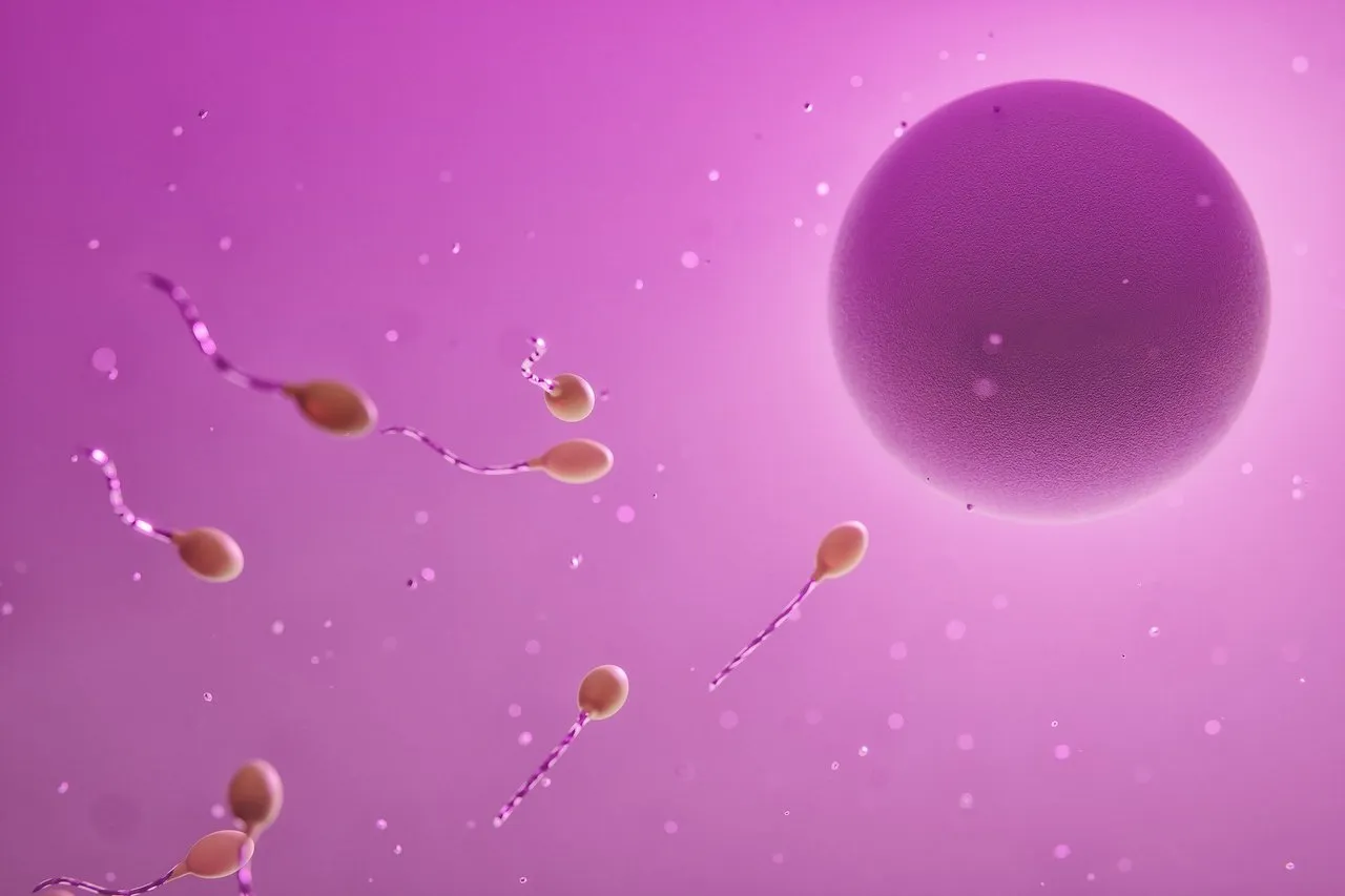 Image of ovum and spermatozoa
Credit: Videomediaart Public domain.
We are born with all the eggs we are going to have and we lose them throughout life.
Hormones produced by the ovaries are very important because they: regulate the menstrual cycle; contribute to female sexual characteristics; participate in pregnancy, childbirth, and breast milk production; contribute to the health of the bones, heart, liver, brain, and other organs; influence mood, sleep, and sexual desire.
Fallopian tubes
They are two muscular ducts located on both sides of the uterus, with an approximate length of 10 cm.
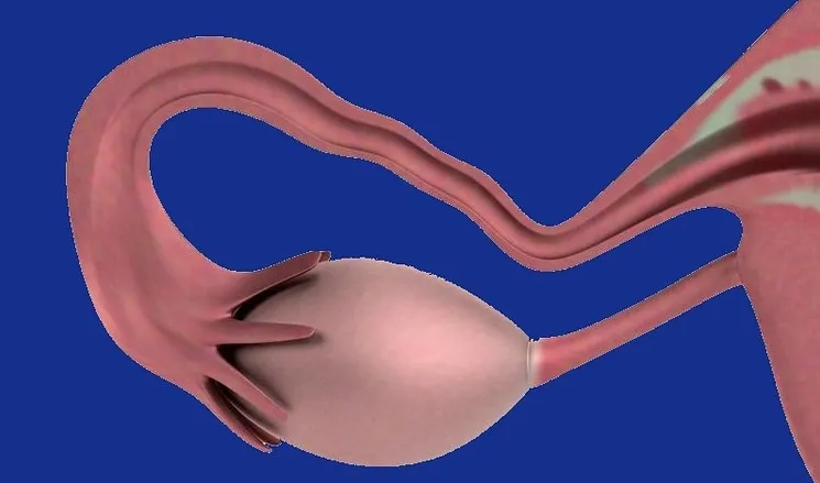 Image of fallopian tube and ovary.
Credit: Therapractice Public domain.Edited in canva
At its distal end, it has finger-like structures, called fimbriae, whose purpose is to surround the ovary. Likewise, in its interior, it has hair-like arrangements called cilia which, when contracted, transport the spermatozoa to the ovum and also transport the ovum (unfertilized) or the embryo (fertilized) to the uterus.
Uterus
It is the largest internal organ, pear-shaped and pear-sized. Its main function is to host the fetus and placenta during pregnancy. It has two differentiated segments, the body, and the cervix.
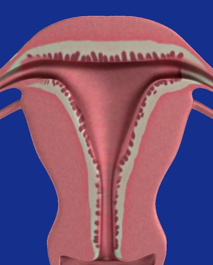 Image of uterus.
Credit: Therapractice Public domain.Edited in canva
The uterine body is made up of two layers:
- Myometrium (thick muscular layer), which expands during pregnancy and contracts during childbirth, these are the so-called uterine contractions.
- Endometrium (inner layer), which varies in thickness throughout the menstrual cycle, thickens after ovulation in expectation of a fertilized egg. If there is no fertilization, the endometrium breaks down, thins, and sheds, which is what we call menstruation.
Cervix
It is a narrow structure located in the lower part of the uterus and connects to the vagina. It has a cylindrical shape and thick walls.
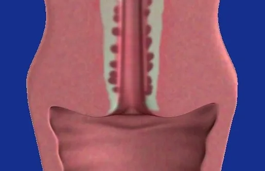
Credit: Therapractice Public domain.Edited in canva
Among its functions is the production of mucus, which prevents sperm from entering the uterus when the woman is pregnant; it protects against bacteria, keeps the vagina healthy, and allows the drainage of fluids, such as menstrual blood.
The cervix, during childbirth, can dilate, opening up to 10 cm wide.
Vagina
It is a flexible muscular canal, varying from 6 to 8 cm in length. It is located anatomically in front of the rectum and behind the bladder and urethra. It connects the internal and external genitalia.
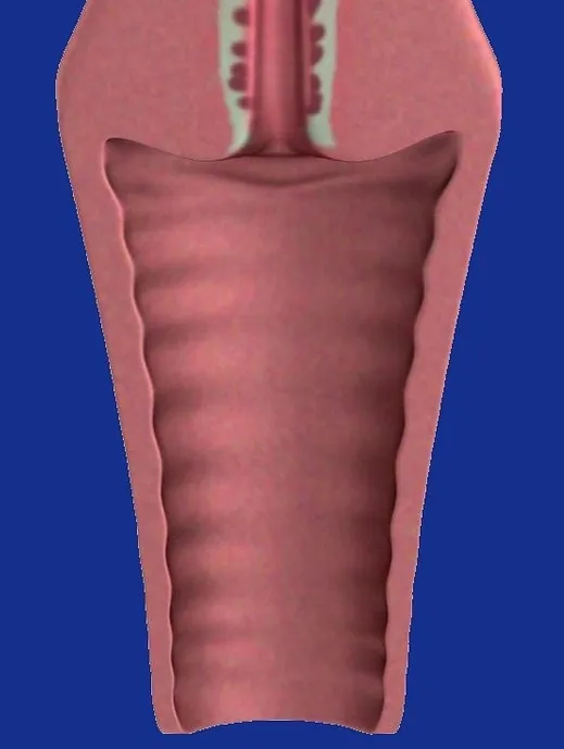
Credit: Therapractice Public domain.Edited in canva
Among its functions are described:
- It houses the penis during penetration and collects sperm from ejaculation which will then travel to the cervix.
- It has glands that produce a substance called vaginal discharge, which lubricates the vagina and its composition varies according to the menstrual cycle.
- Its walls are covered with mucus and harbor Lactobacillus spp, a bacterium that keeps the pH low and prevents the presence of harmful bacteria.
- It widens during childbirth (birth canal) and can also shrink.
- It is the outlet for blood produced during a woman's menstruation.
Final considerations
Practicing self-observation and knowing our body, and its anatomy, is important for the care of our health. This will allow us to detect in time any situation that requires review and specialized assessment.
A visit to a specialist (gynecologist) should be made at least once a year.
Taking care of our health is to love ourselves, value ourselves, strengthen our self-esteem and generate well-being for a full life.

Thus we conclude the first installment of the female anatomy. In the second part, we will discuss the external female genitalia.
Bibliographic references consulted
-Hoare B., Khan Y. Anatomy, Abdomen and Pelvis, Female Internal Genitals. [Internet]. 2022. [quoted 2023 March] Available at
-Biggers, A. Female reproductive organ anatomy. [Internet]. 2021 [quoted 2023 March] Available at
-Cleveland Clinic. Female Reproductive System. [Internet]. 2022. [quoted 2023 March] Available at
-CMG-ATLANTA. Female Anatomy. [Internet] 2020 [quoted 2023 March]. Available at

