Before going into the details of the structure and function of the cochlea, I would only give some hints on other components of the auditory system. The organ of hearing is in fact a particularly complex system and it would be a shame not to say a few words about its other constituents.
The outer ear, at first sight, is little more than a funnel. The "little more than" actually hides subtleties that would be excessive to study here. Suffice it to say that the particular shape of the ear creates variations in the sound signal that the brain uses to figure out if the sound source is behind, above or in front of our head.
The ear canal itself, or external auditory meatus, at first sight, is little more than a pipe, but in reality it is a tuned duct centered on the frequencies of the auditory spectrum, where lies the human voice.
The middle ear appears already to be a more sophisticated structure. The eardrum and the three ossicles transform the oscillations of the air into mechanical oscillations.
The eardrum directly interfaces with the environment, while the three ossicles perform a real impedance adaptation by taking the relatively large vibration of the tympanic membrane and transform them into oscillations of less amplitude but greater pressure, able to put in motion the cochlear mechanism described later.
But the main topic of this article is the cochlea, which is the organ through which these vibrations are converted into nerve impulses, and that is what we will focus on, starting next paragraph.


The scala tympani has an opening on one side called oval window where is connected to the stapes (stirrup). This is the entry point of the sound waves that are transmitted by the ossicular chain to the perilymph. The vibrations are transmitted along the canal of the scala vestibuli up to helicotrema and from there they go to the scala tympani where continue backward until another window, that opens to the middle ear, and is called the round window. The round window is closed by a cartilaginous membrane which allows to cushion the vibrations of the perilymph.

The two types of hair cells have two slightly different functions.
The Outer Hair Cells (OHC) have an active role in the propagation of vibration. In fact, when excited by the vibration of membranes, they contract emitting in turn a vibration proportional to excitation, keeping keeping in vibration the tectorial membrane. This feedback mechanism makes sure that the vibrations, which would normally be damped by the viscosity of the cochlear fluid, are maintained for a longer time by providing a gain of about 100 times (40dB). The mechanical activity of the outer hair cells also produce oscillations that propagate in a retrograde manner until they reache the eardrum. When the eardrum is put into oscillation, are produced the so-called 'otoacoustic emissions', which in recent years have been widely used as a screening method.
The Inner Hair Cells (IHC), represent the real receptor cells, being the only synaptically connected with the fibers of the auditory nerve that carries the acoustic information, transformed into electrical code (action potentials), to the central structures.
What we hear depends on how the mechanical stress of the vibrations affects the Inner Hair Cells. As you can guess from the description of the system, however, the cells do not simply pick up the sound vibrations, that are transmitted from the bracket to the oval window, but they rather react to the vibration of the whole system where they are physically anchored.
So, to understand the functioning of the system, we must first understand what happens when a sound puts in vibration the various membranes of the cochlea.
Having seen how the devices that operate in this manner are common and widespread, we would think that in the cochlea there is not more than the equivalent of an organic microphone that adjusts a nerve signal in proportion to oscillation that is transmitted by the ossicles. Then, as in MP3 players there is a discretization of the signal (that is, a precise numerical value is assigned to the intensity of the signal at a given instant), you may think that the hair cells simply emit discrete discharges in proportion to the intensity of vibration which they are subjected.
This is only partly true. The reality is a bit more complicated. Let's see why.
By convention, we assume that the spectrum of audible by humans extends from 20 to 20,000. Honestly, I doubt that most of the people is really able to hear anything above 16kHz, to be optimistic.
Since our nervous system is made up of neurons, our cochlear transducer is in need of actually make a sampling of the signal, because neurons operate in discrete pulses, but collides with the physiological limit of the neurons themselves, which are able to generate a pulse at intervals to a minimum of a few tenths of a millisecond.
This poses a major limitation to the maximum frequency attainable. In fact, the neurons are able to perform a synthesis of the two effects: the tone coding via the tonotopic arrangement along the basement membrane, of which we speak in the next section, and a time coding using a system of phase lock.
Moreover, even if the inner ear was able to really achieve a sampling of the signal at that frequency, this flow of information, once received by the brain, it should be subjected to a processing extremely onerous. In fact, without imagining a re-conversion of the signal into an analogic one, the brain would be to perform some discrete Fourier transforms in order to filter the signal and extract the information, processing tens of thousands of values per second.
With the available hardware, the nature would not have been able to get a working system in real time in the same way they work today electronic devices.
Then th nature followed a different path: she created a mechanical spectrum analyzer, or electromechanical if you prefer.
The mechanism of operation of the cochlea is not that of a microphone which detects the sound intensity moment by moment and associates some kind of nervous stimulation. It is instead a system that associates specific nervous stimuli to each frequency present in the perceived sound!That is, at a given moment, for example, if we are listening to the sound of a contrabass, there will be some hair cells, dedicated to low frequencies, that will send a strong impulse to the brain, plus some impulse, much less intense, produced by the cells dedicated to the high frequencies, which are excited by the sound of the rubbing of the bow's horsehair across the strings.
That above is the operating principle of our biological spectrum analyzer. In the next section we will see how the nature has achieved this goal.


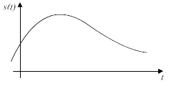
As we said before the waves enter the cochlea through the oval window, which supports the stapes. A pressure wave will produce a deflection of the membrane towards the scala tympani, and vice versa. Due to its structure, however, the basilar membrane will not react the same way along all its length to the various stresses. The change of mass and stiffness along the membrane causes at each point the resonance frequency to be different. In particular, the high frequencies will make vibrate more the portion of the membrane closest to the oval window, while the lower frequencies will make vibrate the more distant portion, nearest to the helicotrema.Thanks to this particular structure of the membrane, the elastic stiffness is not constant, but is about 50000 times larger at the base than the apex decreasing with approximately exponential law.
In the following image you can see the pattern of the resonance frequencies as suggested by Hermann von Helmholtz who is the scholar who first proposed this model.
When a complex sound, consisting of different frequencies, enters the cochlea, the result will be that different points of the basilar membrane will vibrate with different amplitudes in function of the spectral composition of the input signal, stressing in a different way the hair cells which are distributed along the membrane. The fact that the sensitive cells are excited by different tones at different points makes that their distribution is called tonotopic. This organization of nerve fibers is preserved all the way to the brain and the primary auditory cortex too retains a tonotopic structure.