I repeated the transfection of HL-60 cells last week and captured images on the confocal microscope today. The grey brightfield images are on the left. Black and red fluorescent images are on the right. The images on top are of normal HL-60 cells. The images on the bottom are HL-60 cells one week after transfection.
All cells were fixed in 2% paraformaldehyde and mounted with an antifade media. A lot of cellular degradation has occurred in the transfected cells, but you can see the lone intact cell expressing the red fluorescent protein tag in the center of the frame. Untransfected HL-60 cells do not have any red fluorescent protein. All images are 45X and the fluorescent images were obtained using a 552nm laser.
I need to repeat the experiment again with new cells from ATCC. I have been doing all of the preliminary studies on HL-60 cells that were frozen in Liquid Nitrogen (LN2) 20 years ago. It will be a few weeks before my new shipment of plasmid arrives as well. So, until next time my fine fellow geek friends! Eureka!
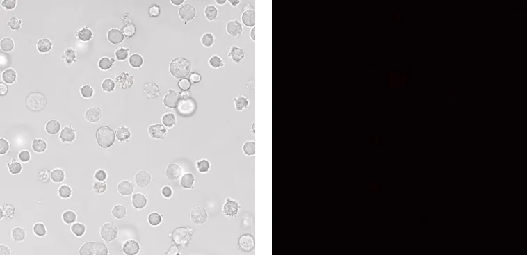
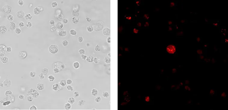
I used the Leica TCS SP8 Confocal Microscope to obtain these images. The SP8 is an inverted laser powered single-point True Confocal Scanning system (TCS) allowing the user to produce publication quality high resolution images of fluorescently labeled histologically fixed tissues and cells, as well as cultured live cells.
