
Hola amigos, un placer estar por acá nuevamente,trayendo información directa y confiable de el área de la medicina, ésta vez, quiero compartir todo lo relacionado con la Laringe, su constitución anatómica especialmente; comencemos pues.
Hello friends, a pleasure to be here again, bringing direct and reliable information from the area of medicine, this time, I want to share everything related to the Larynx, especially its anatomical constitution; let's start then.

La laringe, es una estructura móvil, que forma parte de la vía aérea, actuando normalmente como una válvula que impide el paso de los elementos deglutidos y cuerpos extraños hacia el tracto respiratorio inferior. Además permite el mecanismo de la fonación diseñado específicamente para la producción de la voz. La emisión de sonidos está condicionada al movimiento de las cuerdas vocales. Son los movimientos de los cartílagos de la laringe los que permiten variar el grado de apertura entre las cuerdas y una depresión o una elevación de la estructura laríngea, con lo que varía el tono de los sonidos producidos por el paso del aire a través de ellos. Esto junto a la disposición de los otros elementos de la cavidad oral (labios, lengua y boca) permite determinar los diferentes sonidos que emitimos.
The larynx is a mobile structure, which is part of the airway, normally acting as a valve that prevents the passage of swallowed items and foreign bodies into the lower respiratory tract. It also enables the mechanism of phonation specifically designed for voice production. The emission of sounds is conditioned to the movement of the vocal cords. vocal fold movement. It is the movements of the cartilages of the larynx that allow for the allow varying the degree of opening between the cords and a depression or elevation of the laryngeal structure, thus varying the pitch of the sounds produced by the passage of air through them. This, together with the disposition of the other elements of the oral cavity (lips, tongue and mouth) allows us to determine the different sounds we emit.

La laringe se encuentra situada en la porción anterior del cuello y mide aproximadamente 5 cm de longitud, siendo más corta y cefálica en las mujeres y especialmente en los niños. Se relaciona con los cuerpos vertebrales C3-C6. Su estructura está constituida por un esqueleto cartilaginoso al cual se unen un grupo importante de estructuras musculares y en donde la mucosa adquiere características particulares.
The larynx is located in the anterior portion of the neck and measures approximately 5 cm in length, being shorter and cephalic in females and especially in children. It is related to the vertebral bodies C3-C6. Its structure is constituted by a cartilaginous skeleton to which an important group of muscular structures are joined and where the mucosa acquires particular characteristics.
CARTILAGES OF THE LARYNX
El esqueleto laríngeo está formado por seis cartílagos: Epiglotis, tiroides, aritenoides, corniculados, cuneiformes y cricoides.
Cartílago tiroides:
Cartílago hialino que limita la laringe anterior y lateralmente. Consiste en dos láminas
cuadradas que se fusionan anteriormente en la línea media. Sobre el punto de fusión se
encuentra la escotadura tiroidea. Estas láminas divergen hacia atrás formando un ángulo que en
el hombre es de 90º y en la mujer de 120º. Desde el borde posterior de cada lámina se proyectan
dos cuernos, uno superior y otro inferior. El cuerno superior recibe la inserción del ligamento
tirohioideo lateral. El cuerno inferior se dobla levemente hacia medial y articula en su cara
interna con el cartílago cricoides.Cartílago cricoides:
Cartílago hialino que tiene la forma de un anillo de sello. Se encuentra inferior al cartílago tiroides. Hacia anterior y lateral el anillo se adelgaza formando el arco, pero posteriormente se expande en una lámina gruesa y cuadrada. En la parte superior de la unión del arco con la lámina hacia lateral se encuentra la faceta que articula con el cartílago tiroides. En este mismo
punto hacia superior se encuentra una segunda faceta para la articulación con el cartílago
aritenoides. El cartílago cricoides forma el único anillo cartilaginoso completo del esqueleto
laríngeo, y su preservación es esencial para mantener cerrada la vía aérea.Epiglotis:
Cartílago fibroelástico con forma de hoja que se proyecta hacia arriba detrás de la lengua y el hueso hioides. La delgada porción inferior se inserta a través del ligamento tiroepiglótico al ángulo entre las láminas tiroideas, bajo la escotadura tiroidea. La ancha porción superior se dirige hacia arriba y hacia atrás. Se conecta al hueso hioides por el ligamento hioepiglótico. Su borde superior es libre. En su cara anterior está cubierta por mucosa que viene desde la lengua. En la línea media esta mucosa se eleva para formar el pliegue glosoepiglótico medio y a cada lado de la epiglotis forma los pliegues glosoepigloticos laterales, que pasan hacia la faringe. La depresión que se forma a cada lado del pliegue glosoepiglótico medio se conoce como Vallécula. Desde cada lado de la epiglotis la mucosa se continúa como un pliegue que pasa
hacia los cartílagos aritenoides, y se conoce como pliegue ariepiglótico.Cartílago aritenoides:
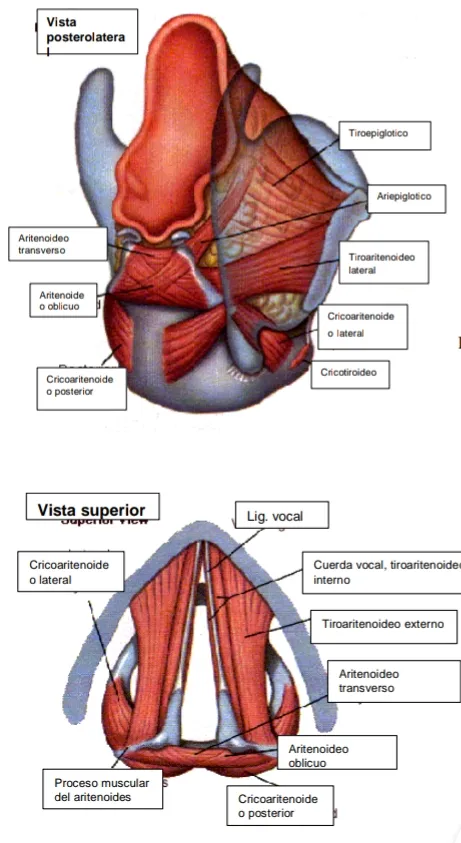
Son dos cartílagos hialinos, de forma piramidal, ubicados sobre el borde superior de la lámina del cartílago cricoides en el borde posterior de la laringe. El vértice se curva hacia atrás y medialmente para la articulación con el cartílago corniculado. El ángulo lateral se prolonga hacia atrás y lateralmente para formar el proceso muscular en el cual se insertan algunas fibras de músculos intrínsecos de la laringe como cricoaritenoideo posterior y cricoaritenoideo lateral. El ángulo anterior se prolonga hacia delante para formar el proceso vocal, al que se inserta el ligamento vocal.Cartílago Corniculado o de Santorini:
Son dos cartílagos fibroelásticos, ubicados por encima del cartílago aritenoides. Dan rigidez a los repliegues ariepiglóticos.Cartílago Cuneiforme o de Wrisberg:
Son dos cartílagos fibroelásticos muy pequeños ubicados a nivel del repliegue ariepiglótico, al
cual también confieren rigidez.
The laryngeal skeleton is formed by six cartilages: epiglottis, thyroid, arytenoids, corniculate, cuneiform and cricoid.
Thyroid cartilage:
Hyaline cartilage that limits the larynx anteriorly and laterally. It consists of two square laminae
fuse anteriorly in the midline. Above the point of fusion is the thyroid notch. These laminae diverge posteriorly forming an angle which in man is 90º and in the male is 90º and in the female 120º. From the posterior edge of each lamina project two horns, one superior and the other superior and the other inferior. The superior horn receives the insertion of the lateral thyrohyoid ligament. The inferior horn bends slightly medially and articulates on its internal aspect with the crico-cranial cartilage on its internal aspect.Cricoid cartilage:
Hyaline cartilage that is shaped like a signet ring. It is inferior to the thyroid cartilage. Anteriorly and laterally the ring thins to form the arch, but posteriorly expands into a thick, square sheet. Superior to the junction of the arch with the lamina laterally is the facet that articulates with the thyroid cartilage. At this same
superiorly there is a second facet for articulation with the arytenoid cartilage. The cricoid cartilage forms the only complete cartilaginous ring of the laryngeal skeleton, and its preservation is essential to keep the airway closed.Epiglottis:
Fibroelastic, leaf-shaped cartilage that projects upward behind the tongue and hyoid bone. The thin lower portion inserts through the thyroepiglottic ligament to the angle between the thyroid laminae, below the thyroid notch. The broad upper portion is directed upward and posteriorly. It is connected to the hyoid bone by the hyoepiglottic ligament. Its superior border is free. On its anterior aspect it is covered by mucosa coming from the tongue. In the midline this mucosa rises to form the medial glossoepiglottic fold and on each side of the epiglottis it forms the lateral glossoepiglottic folds, which pass into the pharynx. The depression formed on each side of the medial glossoepiglottic fold is known as the vallecula. From each side of the epiglottis the mucosa continues as a fold passing into the arytenoid cartilages, and is known as the aryepiglottic fold.Arytenoid cartilage:
These are two hyaline cartilages, pyramidal in shape, located over the upper edge of the lamina of the cricoid cartilage at the posterior border of the larynx. The vertex curves backward and medially for articulation with the corniculate cartilage. The lateral angle extends posteriorly and laterally to form the muscular process into which some fibers of intrinsic laryngeal muscles such as posterior cricoarytenoid and lateral cricoarytenoid insert. The anterior angle extends forward to form the vocal process, to which the vocal ligament inserts.Corniculate or Santorini cartilage:
These are two fibroelastic cartilages, located above the arytenoid cartilage. They give rigidity to the aryepiglottic folds.Cuneiform or Wrisberg's cartilage:
These are two very small fibroelastic cartilages located at the level of the aryepiglottic fold, which also confer rigidity to the aryepiglottic folds.

MEMBRANES AND LIGAMENTS OF THE LARYNX
Ligamentos extrínsecos:
Son aquellos que unen los cartílagos a estructuras adyacentes a los otros cartílagos y además encierran la estructura laríngea.
En orden cefálico-caudal son:
- Membrana tirohioidea. Desde hueso hioides a escotadura tiroidea.
- Membrana hioepiglótica: Delimita con el cartílago tiroides y la vallécula el espacio preepiglótico.
- Ligamento ariepiglótico: Conforma el relieve del vestíbulo.
- Ligamentos tiroepiglóticos: Unen la base de la epiglotis al cartílago tiroides.
- Membrana cricotiroidea: Desde borde superior del cricoides al borde inferior del cartílago tiroides.
- Ligamento cricotraqueal (desde borde inferior del cricoides al primer anillo traqueal).
Ligamentos intrínsecos:
Son aquellos que unen los cartílagos de la laringe entre sí, y juegan un rol importante en la función de este órgano:
- Membrana cuadrangular: Forma el sistema elástico superior de la laringe, se extiende desde los repliegues ariepiglóticos hacia el aritenoides y el borde de la banda
ventricular. - Cono elástico: Sistema elástico inferior que sube desde el cricoides hasta las cuerdas vocales, termina engrosado en la parte de arriba como ligamento vocal.
- Ligamento vocal: Ligamento que ocupa el borde de la cuerda vocal, entre la mucosa y el músculo de la misma.
Extrinsic ligaments:
They are those that join the cartilages to structures adjacent to the other cartilages and also enclose the laryngeal structure.
In cephalic-caudal order they are:
- Thyrohyoid membrane. From hyoid bone to thyroid notch.
- Hyoepiglottic membrane: Delimits with the thyroid cartilage and the vallecula the pre-epiglottic space.
- Aryepiglottic ligament: forms the relief of the vestibule.
- Thyroepiglottic ligaments: join the base of the epiglottis to the thyroid cartilage.
- Cricothyroid membrane: from the superior border of the cricoid to the inferior border of the thyroid cartilage.
- Cricotracheal ligament (from inferior border of cricoid to first tracheal ring).
Intrinsic ligaments:
They are those that join the cartilages of the larynx together, and play an important role in the function of this organ:
- Quadrangular membrane: forms the upper elastic system of the larynx, extending from the aryepiglottic folds toward the arytenoid and the edge of the ventricular band.
ventricular band. - Conus elasticus: lower elastic system that rises from the cricoid to the vocal cords, ends thickened at the top as vocal ligament.
- Vocal ligament: ligament that occupies the edge of the vocal cord, between the mucosa and the vocal cord muscle.

MUSCLES OF THE LARYNX
Los músculos de la laringe son los responsables de la variedad de movimientos de ella. Estos se clasifican en:
Músculos extrínsecos:
Aquellos que se relacionan con los movimientos y fijación de la laringe. Tienen una inserción
en la laringe y otra fuera de ella.
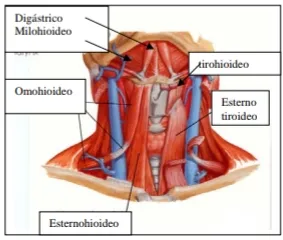
Grupo Depresor de la Laringe
Esternohioideo, Tirohioideo y Homohioideo.
Grupo Elevador de la Laringe
Geniohioideo, Digástrico, Milohioideo, Estilohioideo y Constrictor medio e inferior.
Músculos intrínsecos: aquellos con sus dos inserciones en la laringe, responsables del
movimiento de las cuerdas vocales.
- Músculo Cricotiroideo: se origina de la cara lateral del arco anterior del cartílago cricoides. Algunas fibras se dirigen hacia arriba a la parte posterior del borde inferior de la lámina tiroidea, y otras fibras pasan hacia atrás y lateralmente hacia el cuerno inferior del cartílago tiroides. Es el único músculo de la laringe que es inervado por el nervio
laríngeo superior. Su contracción produce la rotación de la articulación cricotiroidea,
con lo que el tiroides se desplaza hacia abajo y delante, tensando así las cuerdas vocales. - Músculo Cricoaritenoideo posterior: se origina de la superficie posterior de la lámina
del cricoides; las fibras pasan hacia arriba y afuera para insertarse en el proceso
muscular del cartílago aritenoides. Inervado por el N. Laríngeo recurrente. - Músculo Cricoaritenoídeo lateral: se origina en el borde superior de la parte lateral del
arco del cartílago cricoides, sus fibras pasan hacia atrás y arriba para insertarse en el proceso muscular del cartílago aritenoides. Aduce, tensa y alarga las cuerdas vocales.
Inervado por el N. Laríngeo recurrente.
Ambos cricoaritenoideos actuan sinérgicamente en el cierre de la glotis.
Músculo Tiroaritenoideo: se origina de la cara interna de la lámina del cartílago tiroides y de la superficie externa de la membrana cricovocal y se inserta en la superficie anterolateral del cartílago aritenoides. Posee dos porciones: una media (tira vocal) verdadero músculo vocal que en su contracción endurece la cuerda y una porción lateral (tira muscular) la cual forma la masa muscular de la banda y cuya acción cierra el vestíbulo laríngeo. Inervado por
el N. Laríngeo recurrente. - Músculo interaritenoideo: Es el único músculo impar; se encuentra entre los dos aritenoides
y su función es el cierre de la glotis.
The muscles of the larynx are responsible for the variety of movements of the larynx. They are classified as follows:
Extrinsic muscles:
Those that are related to the movements and fixation of the larynx. They have an insertion
They have one insertion in the larynx and another one outside it.
Laryngeal Depressor Group**.
Sternohyoid, Thyrohyoid and Homohyoid.
Laryngeal Elevator Group.
Geniohyoid, digastric, mylohyoid, stylohyoid, and middle and inferior constrictor.
Intrinsic muscles: those with their two insertions in the larynx, responsible for the movement of the vocal cords.
- Cricothyroid muscle: originates from the lateral aspect of the anterior arch of the cricoid cartilage. Some fibers are directed upward to the posterior part of the inferior border of the thyroid lamina, and other fibers pass backward and laterally to the inferior horn of the thyroid cartilage. It is the only muscle of the larynx that is innervated by the superior laryngeal nerve. Its contraction produces rotation of the cricothyroid joint, thereby displacing the thyroid downward and forward, thus tensing the vocal cords.
- Posterior cricoarytenoid muscle: originates from the posterior surface of the lamina of the cricoid; the fibers pass upward and outward to insert into the muscular process of the arytenoid cartilage.
process of the arytenoid cartilage. Innervated by the recurrent laryngeal nerve. - Lateral cricoarytenoid muscle: originates from the superior border of the lateral part of the lateral part of the cricoid cartilage arch.
arch of the cricoid cartilage, its fibers pass backward and upward to insert into the muscular process of the arytenoid cartilage. It adducts, tenses and lengthens the vocal cords.
Innervated by the recurrent laryngeal nerve.
Both cricoarytenoids act synergistically in the closure of the glottis.
Thyroarytenoid muscle: originates from the internal surface of the lamina of the thyroid cartilage and the external surface of the thyroid membrane and inserts on the anterolateral surface of the arytenoid cartilage. It has two portions: a middle one (vocal strip) true vocal muscle that
the vocal cord during its contraction, and a lateral portion (strip muscle) which forms the muscular mass of the band and whose action closes the laryngeal vestibule. Innervated by
the recurrent laryngeal nerve. - Interarytenoid muscle: It is the only odd muscle; it is located between the two arytenoids
and its function is the closure of the glottis.
CLINICAL SUBDIVISIONS OF THE LARYNX
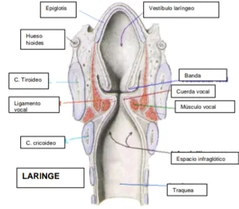
Para describir la patología y semiología laríngea, la laringe puede ser dividida en tres compartimientos, en relación a los pliegues de la mucosa.
Supraglotis, Se extiende desde la punta de la epiglotis a la unión entre el epitelio respiratorio y escamoso en el piso del ventrículo (zona superior de la cuerda vocal).
El vestíbulo laríngeo esta delimitado por la epiglotis y repliegues ariepiglóticos, a ambos lados se continua con las bandas o cuerdas falsas, cuyo borde inferior delimita la entrada al ventrículo
laríngeo. En su parte posteroinferior, el vestíbulo esta delimitado por los cartílagos aritenoides.
Glotis, espacio limitado por la comisura anterior, las cuerdas vocales verdaderas, y la comisura posterior.
Subglotis, desde la unión del epitelio escamoso y respiratorio en la superficie de la cuerda vocal (5mm por debajo del borde libre de la cuerda vocal verdadera) al borde inferior del cartílago cricoides.
To describe laryngeal pathology and semiology, the larynx can be divided into three compartments, in relation to the mucosal folds.
*Supraglottis, extending from the tip of the epiglottis to the junction between the respiratory and squamous epithelium at the floor of the ventricle (upper zone of the vocal cord).
The laryngeal vestibule is delimited by the epiglottis and aryepiglottic folds, on both sides is continued by the false bands or cords, whose lower edge delimits the entrance to the laryngeal ventricle. In its posteroinferior part, the vestibule is delimited by the arytenoid cartilages.
Glotis, space bounded by the anterior commissure, the true vocal cords, and the posterior commissure.
Subglottis, from the junction of the squamous and respiratory epithelium on the surface of the vocal cord (5mm below the free edge of the true vocal cord) to the inferior border of the cricoid cartilage.
INNERVATION OF THE LARYNX
La laringe está inervada por fibras motoras y sensitivas que provienen de los nervios faríngeo superior e inferior de cada lado, los cuatro son ramas del nervio vago. Ambos pares de nervios conducen impulsos aferentes y eferentes y están conectados entre sí por medio de fibras anastomóticas de asociación.
El nervio laríngeo superior:
Es el principal nervio vasomotor, secretor, sensitivo y motor. Tras salir del vago se bifurca en
dos ramas. La externa se dirige hacia abajo para inervar el músculo cricotiroideo. La rama interna atraviesa la membrana tirohioidea para inervar la mucosa de la laringe y epiglotis.
El nervio laríngeo inferior:
Está encargado de la función motora y se separa del vago a diferente nivel en cada lado. En el derecho lo hace a la altura de la arteria subclavia pasando por debajo de ella y ascendiendo por el surco que existe entre la tráquea y el esófago hasta alcanzar el cartílago cricoides, dividiéndose en dos ramas: anterior y posterior.
En el lado izquierdo se separa del vago en el cruce con el cayado aórtico, pasando por debajo de él y ascendiendo hasta la laringe.
Los nervios laríngeos inferiores son independientes, hasta el punto que cada uno puede quedar paralizado separadamente sin que el otro padezca trastornos manifiestos.
The larynx is innervated by motor and sensory fibers from the superior and inferior pharyngeal nerves on each side, all four of which are branches of the vagus nerve. Both pairs of nerves conduct afferent and efferent impulses and are connected to each other by anastomotic association fibers.
*The superior laryngeal nerve:
It is the main vasomotor, secretory, sensory and motor nerve. After leaving the vagus it bifurcates into two branches. The external branch runs downward to innervate the cricothyroid muscle. The internal branch crosses the thyrohyoid membrane to innervate the mucosa of the larynx and epiglottis.
*The inferior laryngeal nerve:
It is in charge of motor function and separates from the vagus at different levels on each side. On the right side it does so at the level of the subclavian artery passing below it and ascending through the groove that exists between the trachea and the esophagus until it reaches the cricoid cartilage, dividing into two branches: anterior and posterior.
On the left side it separates from the vagus at the junction with the aortic arch, passing under it and ascending to the larynx.
The inferior laryngeal nerves are independent, to the point that each one can be paralyzed separately without the other suffering manifest disorders.

IRRIGATION OF THE LARYNX
Las principales arterias que riegan la laringe son: Laríngea superior rama de la Arteria Tiroidea superior que a su vez es rama de la Arteria Carótida externa, y la Arteria Laríngea Inferior rama de la Arteria Tiroidea inferior que es rama del Tronco Tirocervical de la Arteria Subclavia. La Arteria Laríngea superior acompaña a la rama interna del nervio homónimo. Igualmente la Arteria Laríngea inferior acompaña al nervio homónimo.
The main arteries supplying the larynx are: Superior Laryngeal branch of the Superior Thyroid Artery, which in turn is a branch of the external Carotid Artery, and the Inferior Laryngeal Artery branch of the Thyrocervical Trunk of the Subclavian Artery. The Superior Laryngeal Artery accompanies the internal branch of the homonymous nerve.
LYMPHATIC DRAINAGE
La red capilar linfática de la región supraglótica está más o menos separada de la región infraglótica por la escasez de trama linfática en los bordes de los pliegues vocales. La red supraglótica desagua principalmente a los ganglios del grupo cervical superior profundo. La red infraglótica desemboca en los ganglios inferiores del grupo cervical superior profundo.
The lymphatic capillary network of the supraglottic region is more or less separated from the infraglottic region by the scarcity of lymphatic tract at the edges of the vocal folds. The supraglottic network drains mainly to the nodes of the deep upper cervical group. The infraglottic network drains to the inferior nodes of the deep upper cervical group.
Hasta aquí esta pequeña clase magistral, espero aprendan un poco sobre la laringe, importante aparato que nós permite la respiración y, la fonación. Gracias a ella es que hablamos por medio de las cuerdas vocales... Un saludo amigos y espero estar con ustedes muy pronto, trayéndoles novedoso material de primera calidad; Abrazos.
So far this little master class, I hope you learn a little about the larynx, important apparatus that allows us breathing and phonation. Thanks to it is that we speak through the vocal cords .... Greetings friends and I hope to be with you very soon, bringing you new material of first quality; Hugs.

