Hello everyone, it is good to have you around. In my previous post The Anatomy, and Physiology of the Ear, I discussed the anatomy of the ear, which is the sense organ needed for hearing, and maintaining balance. I also mentioned in the post that I will be exploring the sense organ in the body and anything related to it, so in today's post, I will be explaining extensively about the Eyes. The eye is another sense organ of the body, and it is important for sight.
I should state clearly that the post on the Eye will be a series and I will do my best to break down everything you need to know about the eyes, from the External part of the eyes, the Eyeball, then I will explain Phototransduction. I will explain every part of this series carefully and breakdown every necessary thing to be known, to its simplest form.
EYE FACTS
The Anatomy of The Eye
While the eyeball is where most of the Eye teachings start from, if you remember your elementary biology, it is important to know that the eye spreads beyond the eyeball.
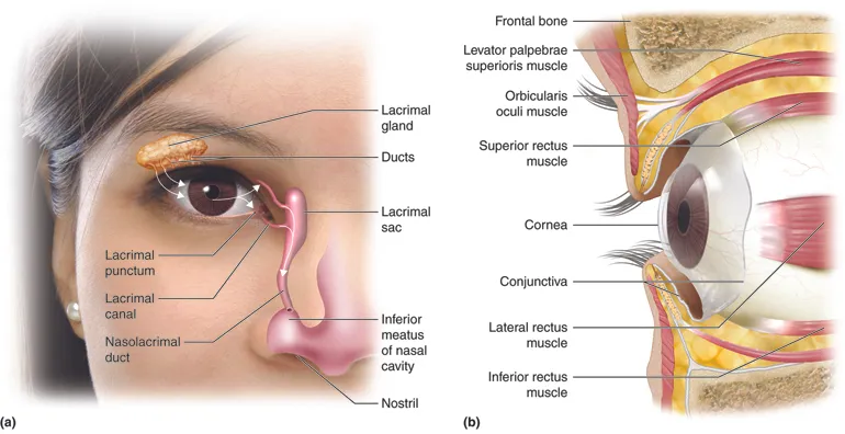
I will be starting with the Palpebral (which you know as the eyelid). This can be categorized into two, the Superior and the Inferior Palpebral. As the name, the superior palpebral is found above, while the inferior palpebral is beneath. The superior Palpebral moves at a greater range compared to the inferior Palpebral. This is as a result of the muscle called the levator palpebrae superioris muscle. The levator palpebrae superioris is innovated by the oculomotor nerve (3rd cranial nerve)..
At the medial distal end of the Palpebral is the Lacrimal coruncle, which is the small reddish mucosal fleshy part. It is made up of sebaceous and sudoriferous glands which secretes whitish substance. Above and below the Lacrimal Coruncle is the Lacrimal Gland or the tear gland, located above the eyeball, at the anterior part of the eye orbit, is integrated by the seventh cranial nerve (facial nerve). The lacrimal gland produces lacrimal fluid, which moves/flows through the cornea for cleansing of the eye surface, protection of the eye surface, and lubricating the surface of the eye. When this fluid runs through the cornea, it is expected that it is channeled out of the eye through a canal called the Lacrimal puncti (Laccrimal Punctum for singular). The Lacrimal puncti are of two types, the inferior Lacrimal punctum (below), and the superior Lacrimal punctum (above). The lacrimal Puncti takes the lacrimal fluid to the lacrimal tubular canaliculi, then moves to the nasolacrimal duct, then into the nasolacrimal sac, after which it gets into the nose through the valve of Hasner..
Underneth the Palpebral is a part of the conjuctiva called the palpebral conjunctiva. The Palpebral Conjuctiva is an underlying conjuctiva that is connected to the upper and lower eyelid. The conjuctiva can be divided into three, one connected to the Sclera known as the bulbar Conjuctiva, another connected to the fornix, known as the forniceal Conjectiva, and the last conjuctiva is connected to the Palpebral which we just discussed.
The Superior Rectus muscle is located around the eye orbit, and connected to the sclera. It is innervated by the 3rd cranial nerve (ocularmotor nerve). The Superior rectus muscle is underneath the levator palpebrae superioris muscle, above the eyeball. The Superior Rectus muscle is important in the eyeball movement. When it contracts, it pulls the eyeball upwards (Superior rotation), and when it retracts, it releases the eyeball to its normal position.. At the side of the eyeball, in the eye orbit, is the another extraocular muscle, known as the Lateral Rectus Muscle which is innervated by the 6th cranial nerve (abducens nerve). This muscle is responsible for abducting the eyeballs, which is rotating the eyes outwardly (laterally).. The Inferior Rectus Muscle is a straight orbital muscle which is responsible for the movement of the eyeball. It is innervated by the 3rd cranial nerve (occularmotor nerve), and rotates the eyeball downwards (depression of the eyeball) during contraction.. The Medial Rectus Muscle is another extraocular muscle innervated by the 3rd cranial nerve. It is located between the medial side wall of the orbit, and the eyeball. It is responsible for rotating the eyeball inwards/Adducting the eyeball..
Inferior to the eyeball is the Inferior Oblique muscle, which is innervated by the 3rd cranial nerve (occularmotor nerve). It is responsible for rotating the eyeball Superiorly and laterally.. Superior to the eyeball, attached to the wall of the orbit medially, and attached to the superior oblique muscle, is the Superior Oblique Muscle. It is responsible for rotating the eyeball downwards internally, and outwards laterally (inferior lateral rotation). The superior oblique muscle is innervated by the 4th Cranial nerves (Trochlear nerve).
The Fibrous Tunic, which is the outer layer of the eyeball, is composed of the Sclera and the Cornea. The Cornea is a clear epithelial tissue which is five layers thick. The Sclera is the white colored surface of the eye. It is a connective tissue to the extraocular muscles, which allows for eyeball movement. The Sclera is covered by the conjunctiva, and is lubricated with Lacrimal fluid which is produced by the Lacrimal gland which we discussed at the upper part of the post.. There are other Tunics in the eye which are the vascular tunic and the Sensory tunic, I will be discussing more on that when we look at the eyeball itself. Currently, we are looking at the ocular surrounding and eye.
Still on the Ocular surrounding is the Optic Nerve. The Optic nerve sends allows for impulses to be sent from the retina to the brain, which then translates whatever is sent. It is easy to say that recognition and interpretation of objects is possible, thanks to the Optic Nerve. The Optic nerve is made up of millions of nerve fibers, and it extends from the posterior part of the eyeball. Optic nerves from the both eyes crosses at the Optic Chiasm/Optic chiasma.. The Optic nerve is located at the lower part of the brain, inferior to the hypothalamus.
Conclusion
In this post, we discussed the anatomy of the eye. In the next post, we will be dissecting the eyeball, and explaining it one after the other, from the Cornea, Pupil, Lens, Retina, Ganglion cell, Aqueous Humor, Ciliary and so on. It is important to know that the eyes isn't just about the eyeball alone, but other muscles, glands, canals and fluids.
Image Reference
Image 1 || Course Hero || Special Senses: Vision