I will officially be launching this programme, with the intent of sharing any interesting findings in the course of conducting clinical diagnosis on patient samples. In essence, once you see the title, it simply will give you an impression of something interesting is coming on board. The pictures shared under the 'results and findings' will all be original pictures of the parasite or whatever microscopic organism or substance seen.
It will be a meaningful read with the one specific objective in mind; making you understand in the simplest of terms, the basic thing about the subject of discourse and how it can either be managed or prevented. With the above specific objective in mind, all the post will run with the following subheadings (introduction and principle, materials and methodology, results and clinical significance / conclusion).
Doing the above will help streamline our discussion and make it straight to the point. Without further adieu, let's begin with our discussion for today.
Introduction
Shistosoma species are parasites that mostly infest humans by piercing and penetrating the skin. They are mainly of five species: Shistosoma haematobium, Shistosoma mekongi, Shistosoma mansoni, Shistosoma intercalatum and Shistosoma japonicum. The infectious and active form of the parasite is the cercariae. They are motile and swim in water and upon contact with human skin, they pierce and penetrate. You might wonder how they do this.
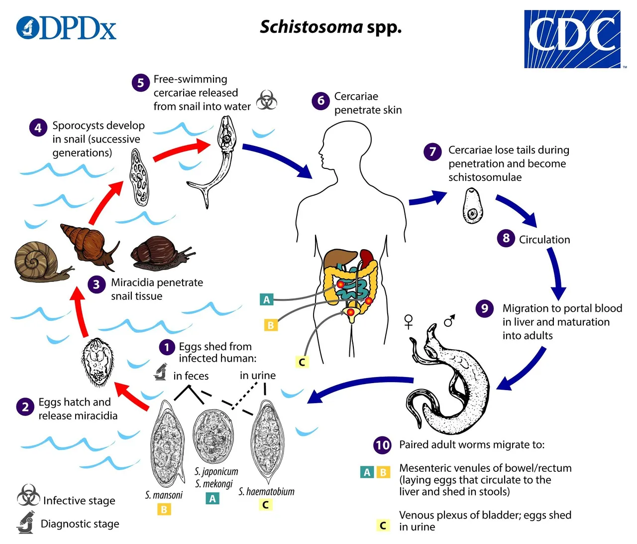
Life cycle of Shistosoma species
First thing you need to understand is that, this parasite are attracted to human skin through the heat, water and electric current when in water. On finding suitable host (humans), they penetrate the skin by secreting proteolytic enzymes (enzymes with the ability of degrading proteins) and they do this penetration with ease.
As they find their way into the body circulation, they in the process lose their tail and form what is Known as the shistosomulae. This is then carried to the liver for maturation into the adult form. Depending on the specie and its predilection site, some move to the rectum or the urinary bladder.
Those found in the bladder are the Shistosoma haematobium. They thrive in the bladder and damage the bladder in the process leading to severe bleeding and blood in the urine. Since they live in the bladder, they also release their eggs in the process and infected patients, when they urinate, pass out this eggs into the environment.
The adult species found in the rectum (Shistosoma mansoni, Shistosoma mekongi, Shistosoma japonicum) release their eggs into the stool and when patient pass out their stools, these eggs are also passed out in the process. These eggs, when they find their way inside water bodies, they hatch and release their larvae (Miracidium) which is motile. Some even hatch while still inside the host body.
The released Miracidium swims around with the help of propellers (flagella) until it finds a suitable water snail. It penetrates the snail's tissues and then forms the next stage of its development - the sporocyst. Snail is important for this parasite to complete it's life cycle.
Releasing from the snail is the infective form of the parasite known as the Cercariae. Just as we earlier explained at the beginning of this introduction. By penetrating another human host, the life cycle continues.
Infection with Shistosoma species like S. haematobium, mansoni and japonicum is common in West Africa and to be specific, Nigeria. Most people that are infected with this parasites are those mostly from the riverine areas and also those that engage in activities related to water (farmers the fisherman). Human presence in water bodies attract the infective form of the parasites when they are present.
The longer humans stay in water that is infested with cercariae, the higher the chances of getting infected with this parasites. It is always advisable to wear protective boots like rain boots and order clothings to prevent penetration by these parasites. Infection with any schistosoma specie is known as Schistosomiasis
Diagnosis
Diagnosis of schistosomiasis in the laboratory is quite simple and less expensive compared to other types of parasites. When infection with schistosomiasis is suspected, the first thing that is requested for is, either these stool or the urine of the patient depending on where the symptoms and signs are noticed.
If it is infection with any of the species, patients most times complain of fever, itchy skin caused by the presence of the eggs released in the body. The major cardinal sign of infection with schistosomiasis especially those associated with the urogenitalia is the presence of blood in the urine of the patient.
The presence of blood in the urine is a sign of the damage caused by the terminal spine of the egg. They use this spine to attach themselves to the walls of the bladder and at the same time causing damaging effects.
To confirm the diagnosis you simply collect the urine or the stool of the patients and examine it under the microscope. For urine sample, all you need to do is to spin or centrifuge the urine sample so as to sediment the eggs. This procedure is also similar with stool sample.
You mix the stool of the patient thoroughly using the turning stick and then take a little portion of the faeces and mix it thoroughly with a physiologic saline. You then spin. Decant the supernatant of both sample and place a drop of the sediments on a clean grease free slide. Cover the slide using a coverslip.
Set up the microscope by focusing using x10 and using x40 objective lens for microscopic evaluation of the parasite. In positive cases that confirms the presence of the parasites, their respective eggs are seen under the microscope. Let's take a look at a real life case we discovered yesterday.
Results and findings
Case of an adult male
The images below are the results from the medical diagnosis carried on the urine sample of an adult (male) who presented with signs and symptoms of infection with the parasites. The mages below were taken using an Android phone.
Taking a shot of parasites through the ocular lens of the microscope can very daunting but still very much possible.
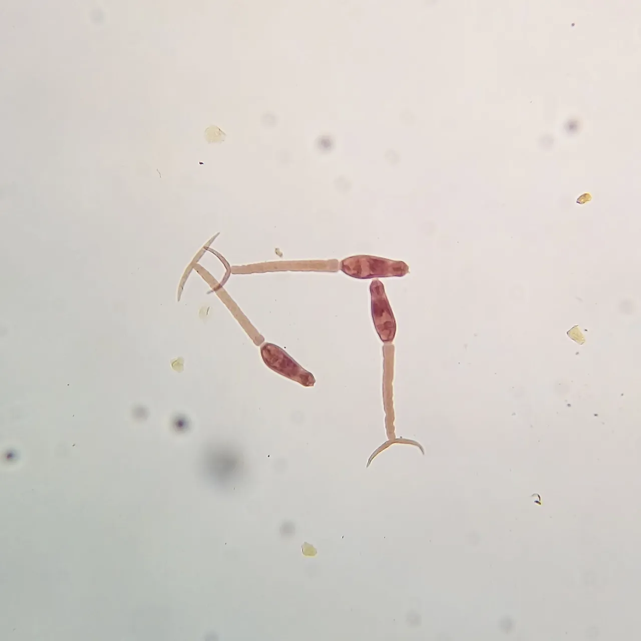
The image above depicts the egg of a matured Shistosoma haematobium found in the urine sample of an adult. An evidence of an active infection with Shistosoma haematobium.
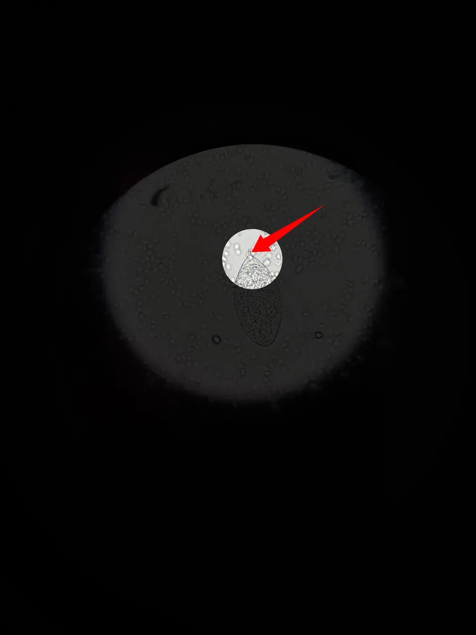
The eggs of Shistosoma haematobium are uniquely identified by the presence of their terminal spine with sharp pointed end. They are mainly found in urine samples. With their spines, they get attached to the walls of the bladder.
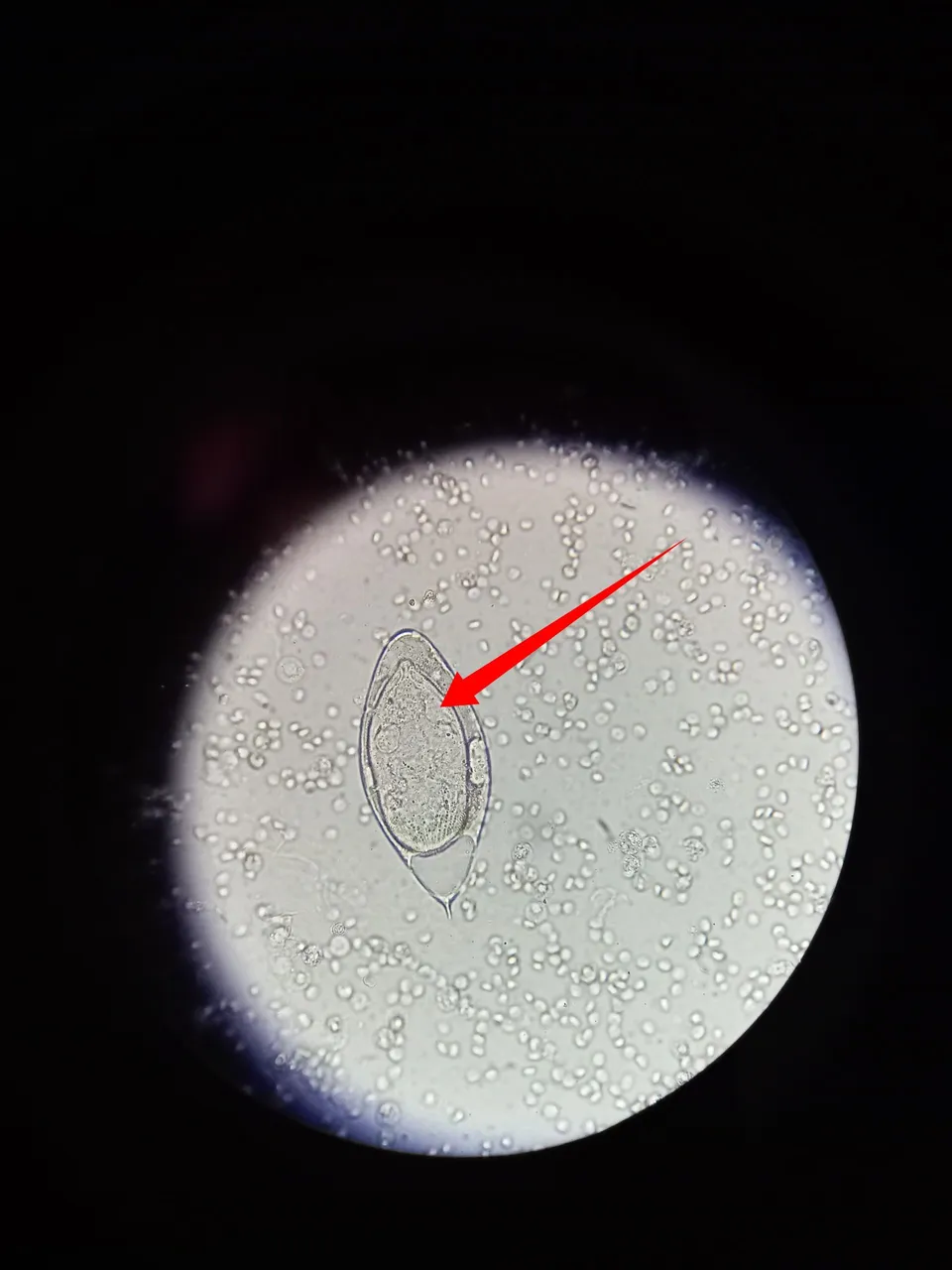
Going further, the matured eggs usually house the Miracidium within them while patiently waiting to be hatched and released once the environment is friendly enough. The above reveals the unhatched eggs containing the Miracidium. The Miracidium is motile in nature and we will reveal this in our experiment in episode two of this laboratory workshop, don't miss it if you want to have a real life view of this parasite.
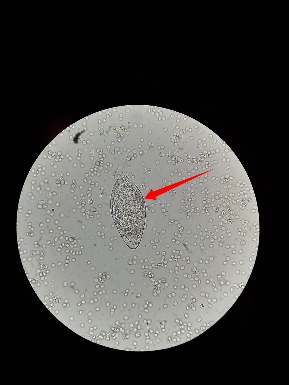
The presence of schistosoma haematobium egg in urine cannot occur without the presence of blood. Most times they go hand-in-hand. Once you suspect or see blood in urine you have to consciously take time to check thoroughly for any possible parasites after you must have ruled out kidney infection.
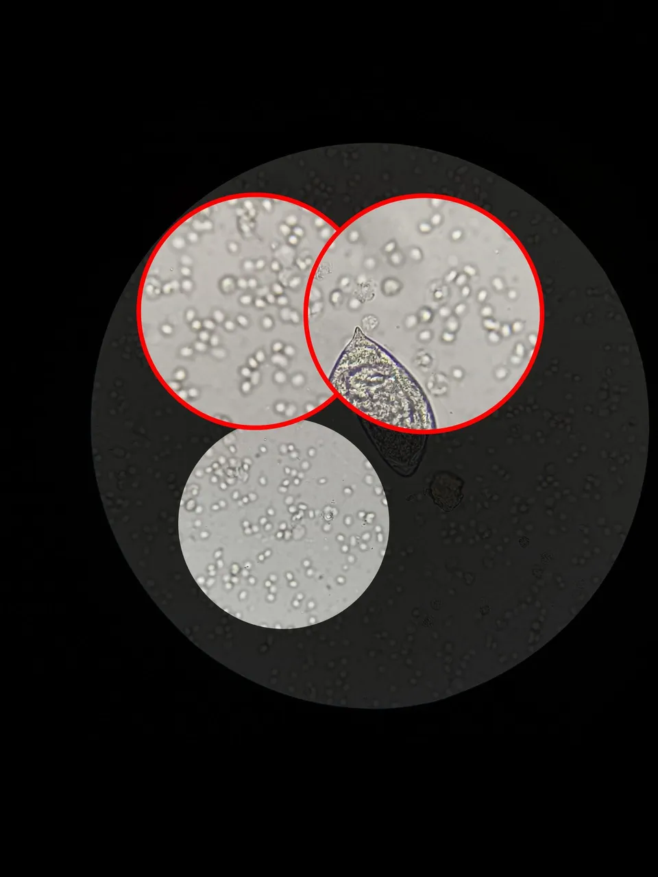
The human blood in urine is always refractive in nature. They are shiny and glistering once present. In the enlarged image above, those shiny round substances are red blood cells in urine. All things being equal, blood is not supposed to be seen in the urine, so when present, it is a clear indication of a pathological issue.
Another important substance you would see in the blood is the presence of pus cells(dead white blood cells). They are larger than normal blood cells and they look granulated.
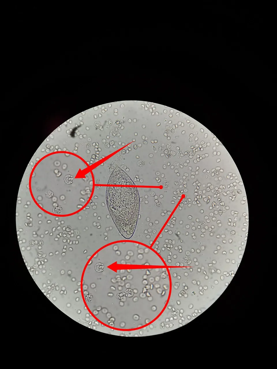
Image of a pus cells in an infected urine sample of a patient with shistosomiasis.
Clinical significance
The presence of schistosoma eggs in urine or stool sample is simply an indication of infection and the disease caused by this parasite as we earlier pointed out is referred to shistosomiasis. Though can be treated, it can also be prevented by simply avoiding long duration in water bodies without clothes since the infective form of the parasite swims freely in water bodies.
Conclusion
Never assume everything is fine once you detect change in colour of your urine most especially when it is red in color. Infection with schistosoma can occur in both males and females hence the need to be very careful.
Please go for treatment once you suspect any abnormal change in your body system. In our next episode we will reveal the motile form (Miracidium) of this parasite once it hatches from the egg and as it swims in the pool of blood cells found in the urine. Stick around!
References
•Parasites
•Facts sheets - Shistosomias
•Mechanisms of Skin Penetration by Schistosoma Mansoni Cercariae