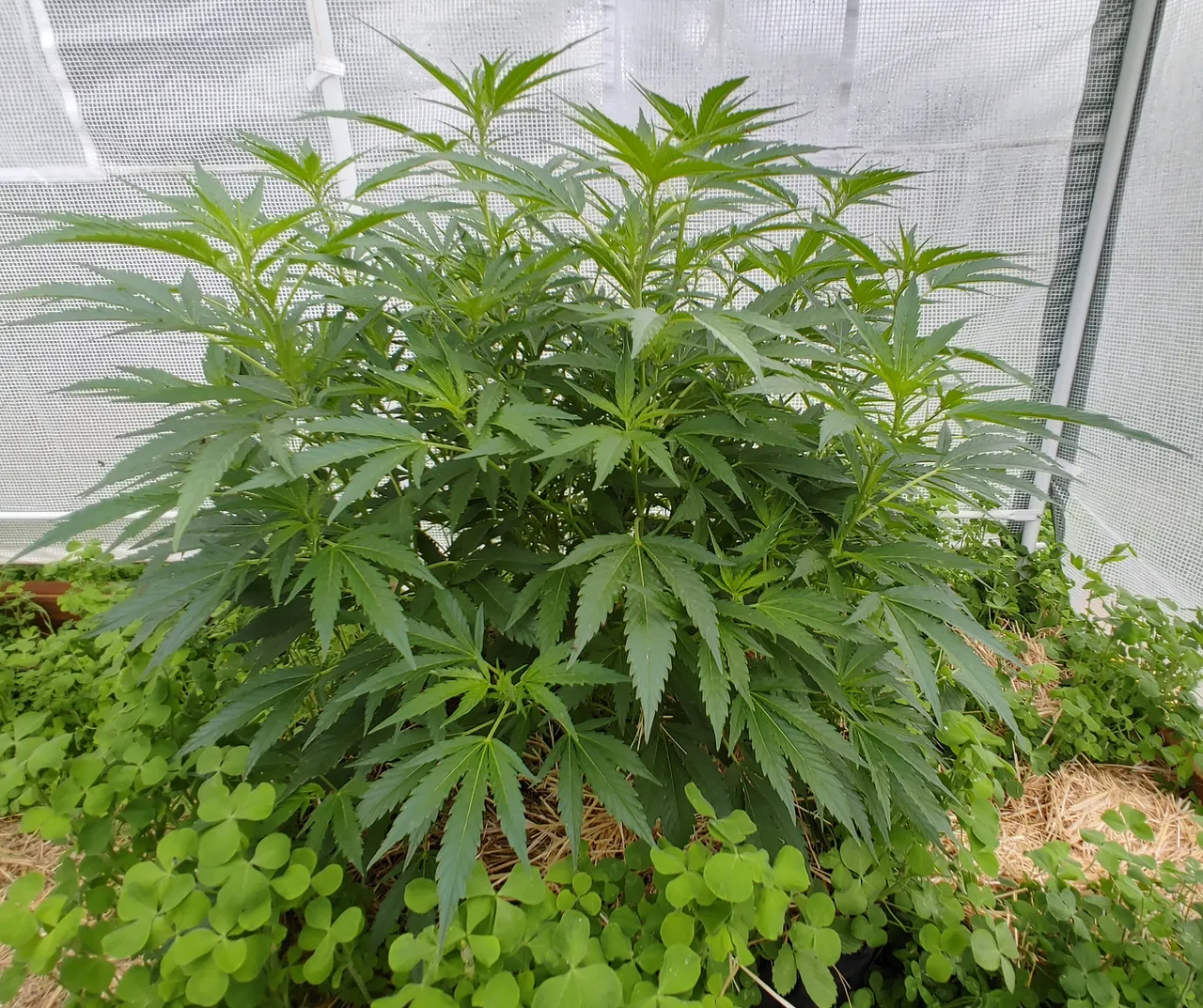
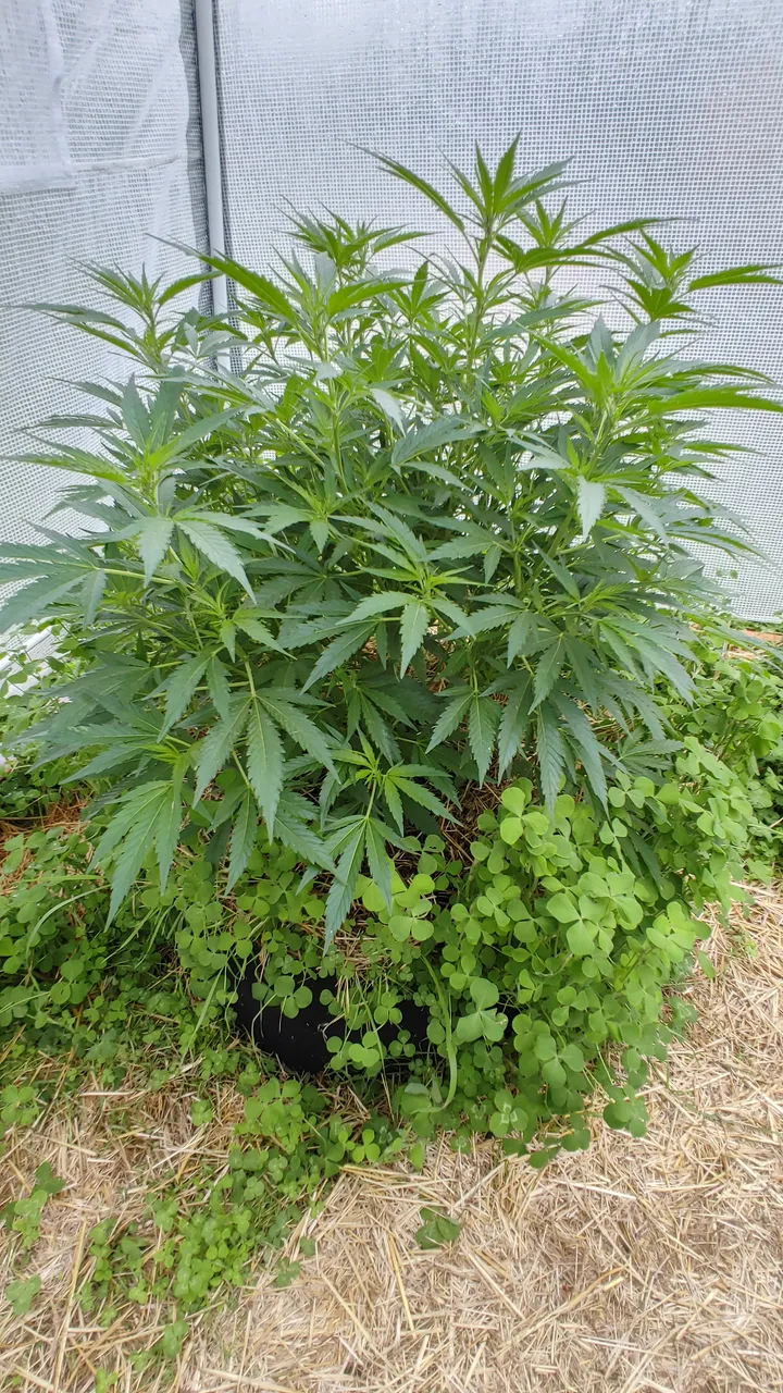
Using a three inch apple corer, I took 3 core samples that were halfway in-between the edge of the fabric pot and the stalk of the plant. I placed the samples in a Ziplock bag and brought it back to my room/lab in order to prepare a sample.
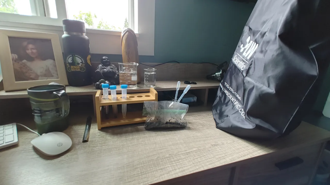
I used a dilution of 10 and two drops on the slide for my sample. First, I scanned for Nematodes at 100x total objective.
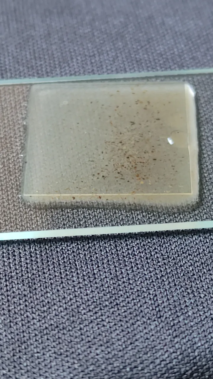
Nematode Scan
Next, using the same sample, I did a main assessment where I looked for filamentous organisms and protozoas at a 400x total magnification.
Starting at the top right area of the 18x18 coverslip and going from left to right in a straight line, I chose 5 random areas in that lane to read the different Fields of View in each area.
The following videos are Five readings in consecutive order.
Reading 1
Reading 2
Reading 3
Reading 4
Reading 5
Now that I did my main assessment and scanned for nematodes, it is time to make a new slide just for my bacterial counts. I will dilute this sample to a dilution factor of 1000 and use 2 drops on the slide.
In five different areas, much like the 5 side of a dice, I will record 5 different areas on the cover slip to count for bacteria. Since there was lots of bacteria count I counted a 1/4 of the screen/cover slip area at 400x total magnification.
Bacterial Count Readings
Results
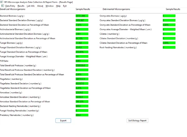
While I did not really see any fungi to write to home about, I did see a crapload of flagellates and bacterial feeding nematodes. Which is a really good thing. I did see one ciliate on my nematode scan which is still concerning. The bountiful amount of flagellates is apparently signs the soil might be over watered as flagellates favor aquatic environments over soil but still are great nutrient cyclers. The ciliate is concerning but watering has been tricky as I can only water once a week, too wet, as well as there being a compaction layer under each fabric container. Do remember that this grow is 20 feet away from a creek so perhaps the extra flagellates are also feature of growing in this area.
So a really good reading in my opinion. Seems my soil here has a lot of nutrient cycling going on!
Are you curious about the microscope and using one for soil analysis? Check out my Soil Food Web mentor Wes in a recent Soil Food Web video about using the microscope!