
Greetings @lifestyle community! It is a pleasure for me to share with you my first post for this great community. This post has a great sentimental value for me since it is essentially about a part of my identity as a person and professional, science. And in this context, these lines are dedicated to one of the research centers where I have trained as a scientist and where I now transmit my knowledge to the new generations. Let's get started!
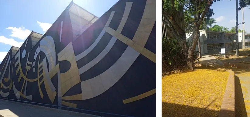
In the city of Caracas, in the spaces of the University City, declared by UNESCO as a World Heritage Site, is the Faculty of Sciences of the Central University of Venezuela. Created in 1958, it is a 3rd and 4th level Study Center with 66 years of experience in the training of human capital for Venezuela. It is made up of 5 schools: Chemistry, Physics, Mathematics, Computing, Geochemistry and the one we are going to talk about today: the School of Biology.
The School of Biology, although it dates back to 1946, was formally created as an institution attached to the Faculty of Sciences in 1958. It is currently made up of 5 departments: Departments: Cellular Biology, Botany, Ecology, Food Technology and Zoology. Each of these departments carries out research activities and 4th level studies respectively.

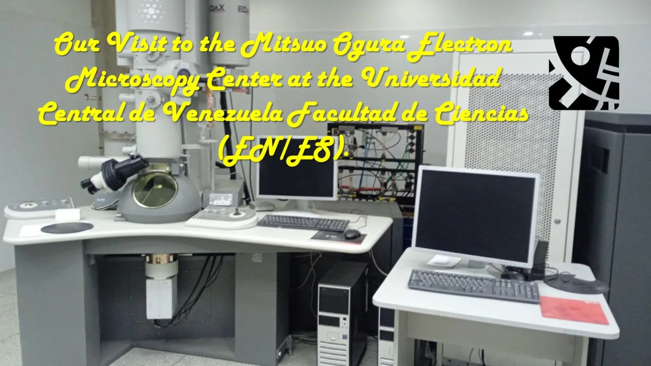
The Institutes of Zoology and Tropical Ecology (IZET), Institute of Experimental Biology (IBE) and Institute of Food Technology are the 3 large institutes dedicated to research and undergraduate and graduate studies. However, there is a Center that is as exceptional as it is full of great historical weight in the School of Biology and it is the one we are going to talk about today: The Mitsuo Ogura Electron Microscopy Center.

Electron microscopy in Venezuela cannot be understood without the participation of the distinguished "Citizen of the World" Humberto Fernández Morán Villalobos. Born in 1924 in the State of Zulia, at the age of 15 he entered the University of Munich to study medicine during WWII. In 1946 in Stockholm he studied Neuroscience and Biophysics which led him to study electron microscopy and conceive cryomicroscopy. His contributions earned him the title of Knight of the Grand Cross of the Royal Order of the Polar Star awarded by King Gustav Adolf of Sweden. From there he returned to Venezuela and founded what would later become the Venezuelan Institute for Scientific Research (IVIC).
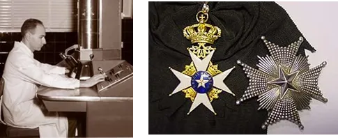
Humberto Fernandez Morán & title of Knight of the Grand Cross of the Royal Order of the Polar Star
Dr. Mitsuo Ogura (1928-200) received his PhD in Medicine from the University of Kyoto, Japan and had dedicated his specialization to the use of electron microscopy within the area of cellular biology. Hired by the Department of Biochemistry and Biophysics of the School of Biology of the Faculty of Sciences, he was one of the most brilliant emigrants who arrived in Venezuela. In 1963 he founded the Electron Microscopy Laboratory in the IBE spaces to finally become the Electron Microscopy Center in 1977 in the spaces of the Faculty of Sciences.
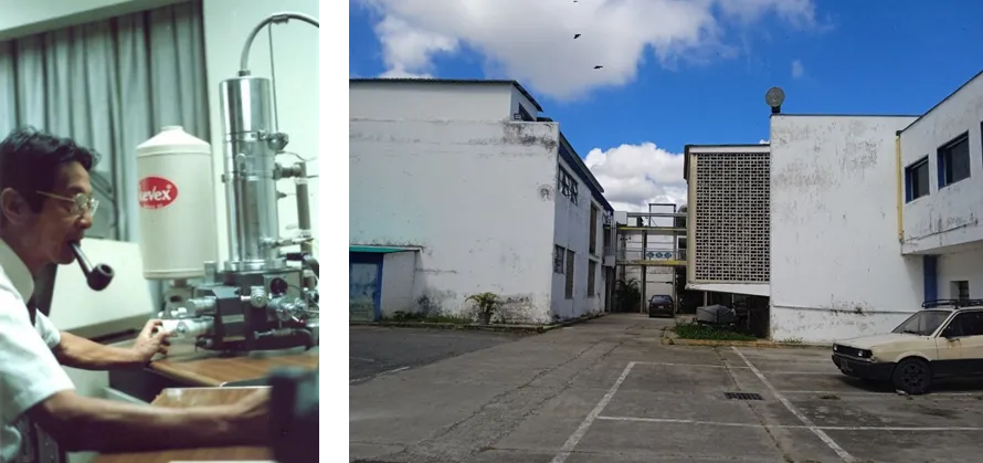
Dr. Mitsuo Ogura (1928-2000) & Initial location of the Electron Microscopy Laboratory (IBE)

The contribution to the creation of the Electron Microscopy Center has been one of the greatest achievements of the School of Biology, since over time it has become a multidisciplinary center and in this context as Head of the Chair of the Cellular Biology Teaching Unit it is my duty to take undergraduate students to know this space of scientific development.
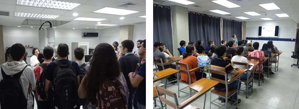
During this visit to the Electron Microscopy Center, students are shown the Old Center, which has virtually become a museum where they can see the first models of microscopes, and then the new center, which has been built to house the new equipment for better maintenance. However, it is important to ask ourselves: What is electron microscopy?
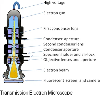
Unlike a classic optical microscope where the microscopic image we observe is the light focused on a sample and collected by high magnification lenses, in the case of a transmission electron microscope the image generated is the product of a bombardment of electrons emitted and concentrated by magnetic capacitors that, when they hit a biological sample, are collected by specific detectors, generating an image with a resolution and magnification thousands of times greater than an optical microscope.
In general, there are two types of microscopes: transmission and scanning. While the former allows you to see through a biological sample, the latter allows you to study its surface. In both cases, they are devices for the ultrastructural study of tissues and cells.
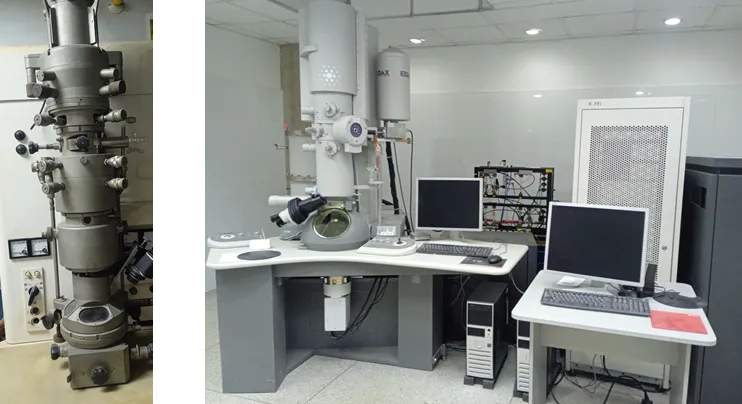
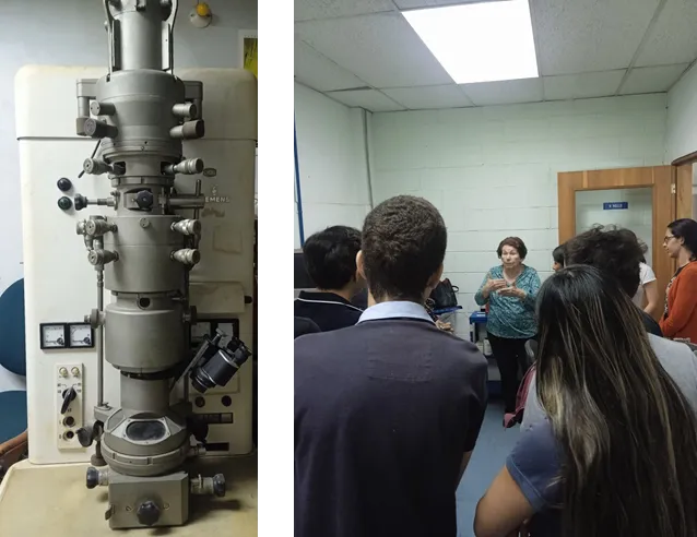
The old Centre houses many of the early pieces of equipment that were used even when it was known as the Electron Microscopy Laboratory. The most notable is one of the first electron microscopes to arrive at the Central University, a Siemens Elmiskop 1, while on the right we can see Prof. Frecculi Dager, Head of the Laboratory of Parasite Biochemistry and one of the last professors to have operated this equipment.
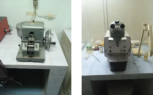
Although it is an art to operate this equipment, it is also an art to prepare the biological samples that will be seen in the electron microscope, this means that ultra-thin sections must be made of the sample to be studied. In the image below we see on the left what the first ultramicrotomes were like and on the right one of the newest equipment, which has a microscope to make the sections.
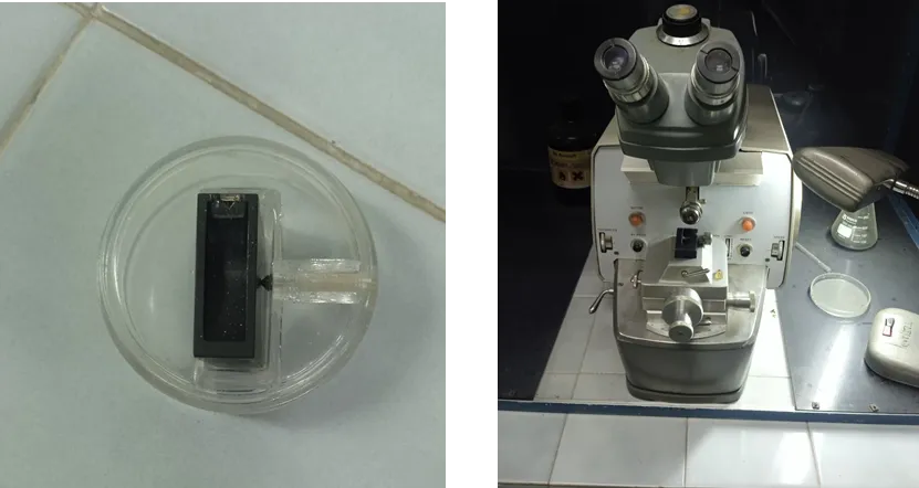
The main problem with making ultra-thin cuts for study in electron microscopy was the fragility and the artifacts that these generated, since at that scale any procedure resulted in the loss of the biological preparation. It is there where Humberto Fernandez Morán shone with his own light by devising the diamond blade to achieve perfect cuts of the samples. In the image you can see the legacy of this exceptional Venezuelan and his mount in the ultramicrotome.
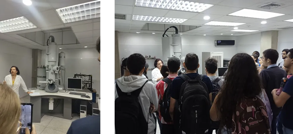
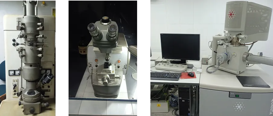
Despite the great economic, social and political turbulence that we are experiencing, the Central University of Venezuela has managed to maintain this important research center, and so currently the Electron Microscopy Center has 2 of the newest electron microscopes, one scanning and one transmission, where we can see Prof. Caribay Urbina explaining the functions of this equipment.
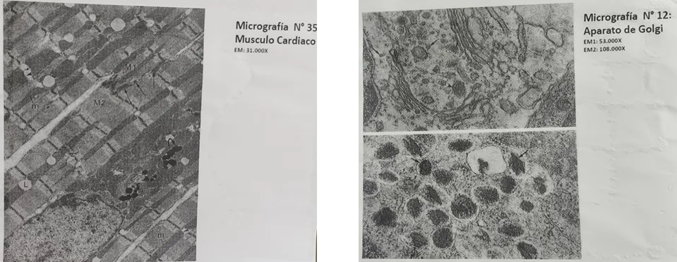
The images obtained are always in black and white and in this case they are images obtained with the transmission electron microscope. The left image shows us what the fibers of the cardiac skeletal muscle look like and the right image shows the Golgi apparatus. As you can see, the degree of resolution of this equipment is significantly high.
This visit has two purposes: the first is to train them as biologists, allowing them to learn about the relationship between structure and ultrastructure and cellular function and how, from here, they are integrated into a complex relationship of systems and tissues. The second aspect is to attract new students to begin a career in the area of ultrastructure, which is a way of keeping the flame of Drs. Fernández and Ogura alive.
And well, dear friends, this was all for the visit that I coordinated for my undergraduate Biology students. They are already more than halfway through their degree and taking them to see this center has great value for me, not only for the academic part but also for the historical part. My main mentors came from this center and have made me the researcher I am today, so the best thing I can do to honor their memory is to transmit my knowledge to the new generations. See you in a next post, best regards!
Saludos comunidad de @lifestyle! Es un gusto para mi compartir con ustedes mi primer post para esta gran comunidad. Este post tiene un gran valor sentimental para mi ya que en esencia se trata de una parte de mi identidad como persona y profesional, la ciencia. Y en este contexto estas líneas están dedicadas a uno de los centros de investigación donde me he formado como científico y en el que ahora trasmito mis conocimientos a las nuevas generaciones. Comencemos!
En la ciudad de Caracas, en los espacios de la Ciudad Universitaria, declarada por la UNESCO como Patrimonio de la Humanidad se encuentra la Facultad de Ciencias de la Universidad Central de Venezuela creada en 1958 es un Centro de Estudios de 3er y 4to nivel con ya 66 años en la formación de capital humano para Venezuela. se conforma de 5 escuelas: Química, Física, Matemáticas, Computación, Geoquímica y de la que vamos a hablar hoy: la escuela de Biología.
La escuela de Biología aunque tiene su origen en los años 1946, su creación formal tiene su creación formal como institución adscrita a la Facultad de Ciencias en 1958. Actualmente se compone de 5 departamentos: Departamentos: Biología Celular, Botánica, Ecología, Tecnología de Alimentos y Zoología. Cada uno de estos departamentos lleva acabo actividades de investigación y estudios de 4to nivel respectivamente.
Los Institutos de Zoología y Ecología Tropical (IZET), Instituto de Biología Experimental (IBE) e Instituto de Tecnología de Alimentos son los 3 grandes institutos dedicados a la investigación y estudios de Pregrado y Postgrado. Sin embargo existe un Centro tan excepcional como lleno de un gran peso histórico en la escuela de Biología y es del que vamos hablar hoy: El Centro de Microscopía Electrónica Mitsuo Ogura.
La microscopía electrónica en Venezuela no puede entenderse sin la participación que tuvo el insigne "Ciudadano del Mundo" Humberto Fernández Morán Villalobos. Nacido en 1924 en el Estado Zulia, a los 15 años ingresó a la Universidad de Münich para estudiar medicina durante WWII. En 1946 en Estocolmo estudia Neurociencia y Biofísica que lo llevo a estudiar microscopía electrónica y concebir la crio microscopía, sus aportes le valieron el Título de Caballero de la Gran Cruz de la Orden Real de la Estrella Polar otorgada por el Rey Gustavo Adolfo de Suecia. De alli vuelve a Venezuela y Funda lo que posteriormente sería El Instituto Venezolano de Investigaciones Científicas (IVIC).
Humberto Fernandez Morán & Título de Caballero de la Gran Cruz de la Orden Real de la Estrella Polar otorgada por el Rey Gustavo Adolfo de Suecia
El Dr. Mitsuo Ogura (1928-200) Dr. en Medicina por la Universidad de Kioto, Japón y había dedicado su especialización en el uso de la microscopía electrónica dentro del área de la biología celular. Contratado por el departamento de Bioquímica y Biofísica de la Escuela de Biología de la Facultad de Ciencias fue uno de los emigrantes más brillantes que llego a Venezuela, en 1963 funda el Laboratorio de Microscopía Electrónica en los espacios del IBE para finalmente en 1977 convertirse en el Centro de Microscopía Electrónica en los espacios de la Facultad de Ciencias.
Dr. Mitsuo Ogura (1928-2000) & Lugar inicial del Laboratorio de Microscopía Electrónica (IBE)
El aporte a la creación del Centro de Microscopía Electrónica ha sido uno de los mayores logros alcanzados por la escuela de Biología, ya que con el tiempo se ha convertido en un centro multidisciplinario y en este contexto como Jefe de Cátedra de la Unidad Docente de Biología Celular es mi deber llevar a los estudiantes de Pregrado a conocer este espacio de desarrollo científico.
En esta visita al Centro de Microscopía Electrónica se les muestran a los estudiantes inicialmente como era el Centro Antiguo que virtualmente se ha convertido en un museo donde pueden contemplar los primeros modelos de microscopios y posteriormente el centro nuevo que ha sido construido para albergar los nuevos equipos para su mejor mantenimiento. Sin embargo es importante preguntarnos: ¿Qué es la microscopía electrónica?
A diferencia de un clásico microscopio óptico donde la imagen microscópica que observamos es la luz enfocada en una muestra y recogida por lentes de gran aumento, en el caso de un microscopio electrónico de transmisión la imagen que se genera es producto de un bombardeo de electrones emitidos y concentrados por condensadores magnéticos que al incidir en una muestra biológica son recogidos por detectores específicos generando una imagen con una resolución y aumento miles de veces mayor que un microscopio óptico.
En general existen 2 formas de microscopios el de transmisión y el de barrido, mientras el primero permite ver a través de una muestra biológica, el segundo permite estudiar su superficie. En ambos casos son equipos para el estudio ultraestructural de tejidos y células.
En el antiguo Centro se ubican muchos de los primeros equipos que fueron utilizados incluso cuando era conocido como Laboratorio de Microscopía Electrónica. El más destacado es uno de los primeros microscopios electrónicos que llegó a la Universidad Central, se trata de un Siemens Elmiskop 1, mientras que en la imagen derecha podemos observar a la Prof. Frecculi Dager Jefa del Laboratorio de Bioquímica de Parásitos y una de las últimas profesoras en haber operado este equipo.
Si bien es un arte operar estos equipos, también lo es preparar las muestras biológicas que se verán en el microscopio electrónico, esto significa que deben hacerse cortes ultra finos de la muestra que se va a estudiar, en la imagen abajo vemos a la izquierda como eran los primeros ultra micrótomos y a la derecha uno de los equipos más nuevos, el cual cuenta con microscopio para realizar los cortes.
El principal problema de realizar cortes ultra finos para su estudio en la microscopia electrónica era la fragilidad y los artefactos que estos generaban ya que a esa escala cualquier procedimiento resultaba en en la pérdida de la preparación biológica. Es alli donde Humberto Fernandez Morán Brilló con luz propia ideando la cuchilla de diamante para lograr cotes perfectos de las muestras, en la imagen puedes ver el legado de este excepcional venezolano y su montura en el ultra micrótomo.
A pesar de la gran turbulencia económica, social y política que estamos viviendo, la Universidad Central de Venezuela ha logrado mantener este importante centro de investigación es así como actualmente el Centro de Microscopía Electrónica tiene 2 de los más nuevos microscopios electrónicos, uno de barrido y otro de Transmisión que es donde podemos observar a la Prof. Caribay Urbina explicarnos las funciones de este equipo.
Las imágenes obtenidas son siempre en blanco y negro y en este caso son imágenes obtenidas con el microscopio electrónico de transmisión, la imagen izquierda nos muestra como son las fibras del musculo esquelético cardíaco y la imagen derecha el aparato de golgi, como pueden ver el grado de resolución de estos equipos es significativamente alto.
Esta visita tiene 2 propósitos, la primera en su formación como biólogos, les permite conocer la relación que tiene la estructura y la ultraestructural con la función celular y como a partir de aquí se integra en una compleja relación de sistemas y tejidos. Como segundo aspecto es atraer a los nuevos estudiantes para que comiencen una carrera en el área de la ultra estructura, es una manera de mantener la llama viva de los Doctores Fernández y Ogura.
Y bien estimados amigos, esto fue todo en la visita que coordiné a mis estudiantes del pregrado de Biología, ya están a más de la mitad de la carrera y llevarlos a conocer este centro tiene un gran valor para mí no solo por la parte académica sino también por la histórica. Mis principales mentores salieron de este centro y me han hecho el investigador que soy ahora así que lo mejor que puedo hacer para honrar su memoria es transmitiéndole mis conocimiento a las nuevas generaciones. Nos vemos en un próximo post, saludos!
Las imágenes fueron tomadas con mi celular (Redmi 9c) La traducción se realizó con la herramienta Deepl
The images were taken with my cell phone (Redmi 9c) The translation was done with the tool Deepl