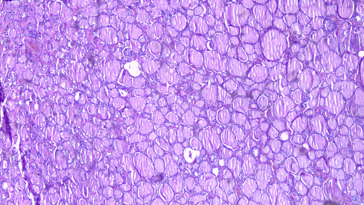
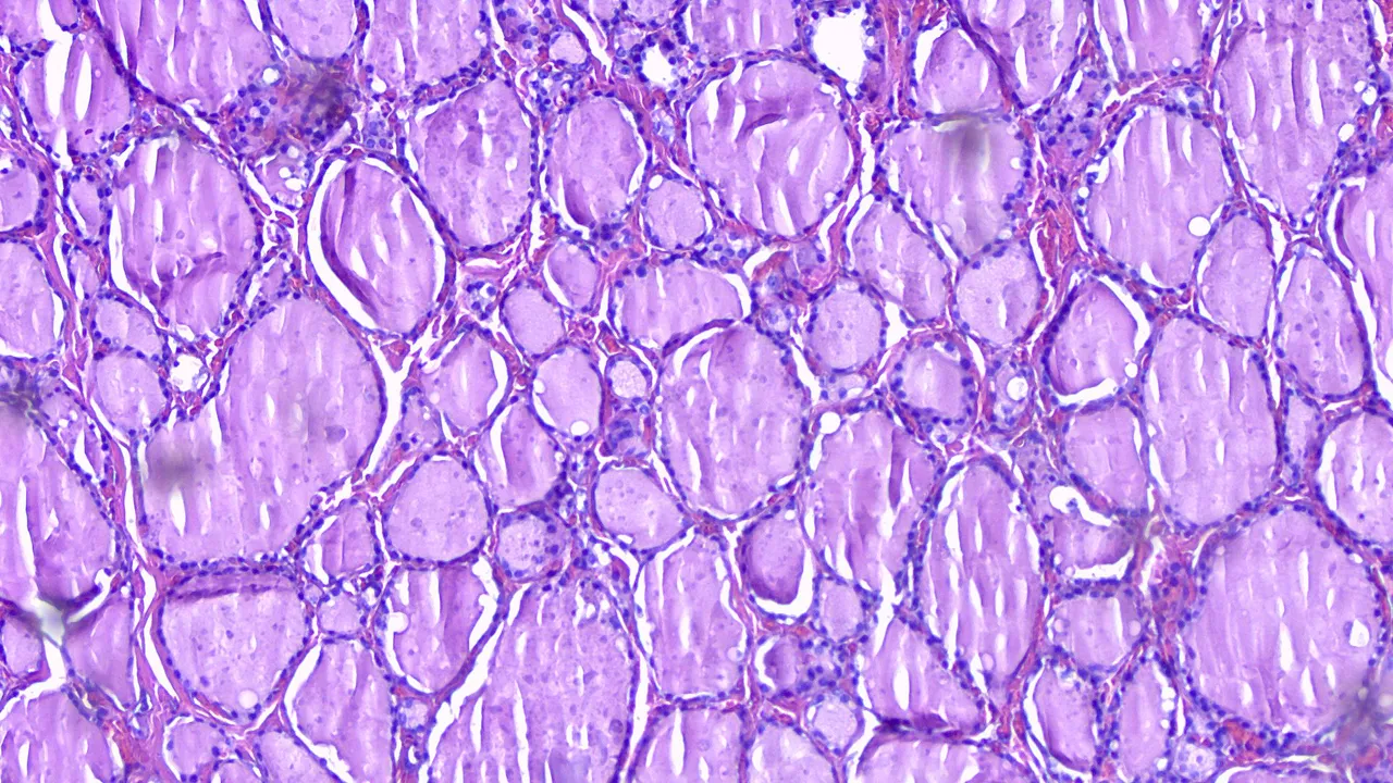
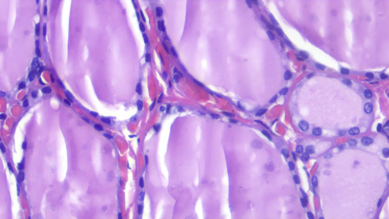
The following three images are the normal architecture of the thyroid gland and then followed up by a case of papillary thyroid carcinoma. Thyroid tissue is made up follicles containing colloid and lined by simple cuboidal epithelium. If you recall your biology class, cuboidal cells are cuboids in shape and the form associated with glands. The cells have round, normochromatic nuclei with inconspicuous nucleoli and moderate cytoplasm.
Taken at Scanner View (40x)
Taken at Low Power View (100x)
Taken at High Power View (400x)
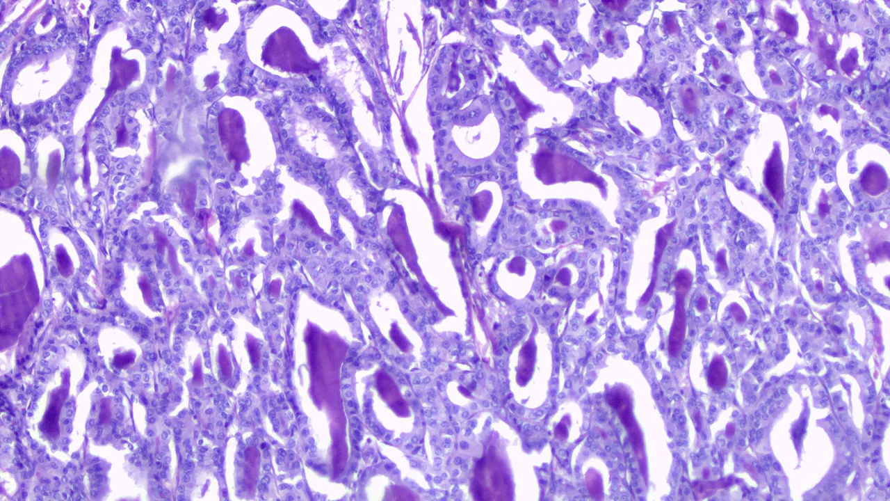
Diagnosis of Papillary Thyroid Carcinoma (PTC) depends on the nuclear features rather than overall architecture. PTC has a lot of histologic variants and some have variable prognosis that are associated with worse prognosis like Tall-Cell variants but I won’t be dwelling on that.
The tumor cells are described as optically clear nuclei (Orphan Annie Nuclei) that can overlap with each other, nuclear grooving and has pseudoinclusions as classic features. Seeing pseudoinclusions raises the confidence that it’s PTC but sometimes you don’t get to see these features all the time. Whenever I see nuclear grooving, optical clearing and overlapping.
Taken at Scanner View (40x)
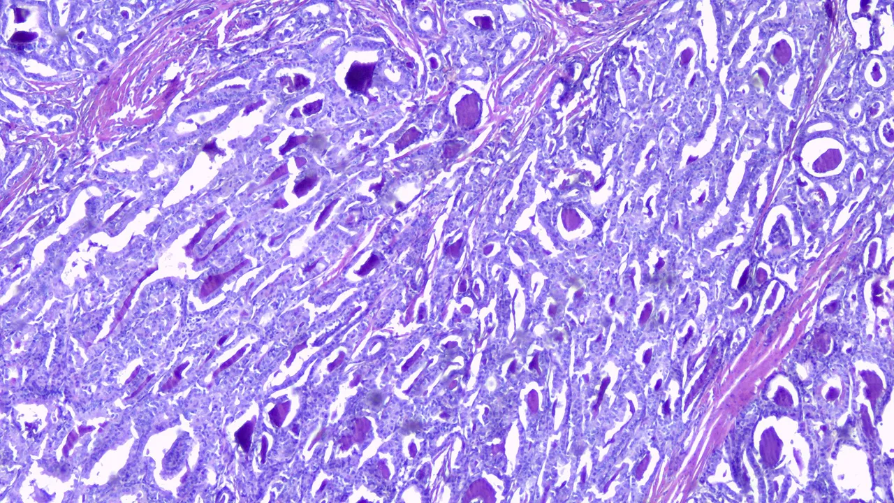
Taken at Low Power View (100x)
Taken at High Power View (400x)
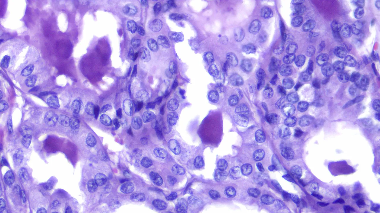
Malignant cells don’t respect personal space that’s why they overlap. Some look like round cells being folded longitudinally (nuclear grooving), and the chromatin inside the nuclei can be optically clear (Orphan-Annie). The staging and prognosis is favorable for people less than 55 years of age and prompt treatment.
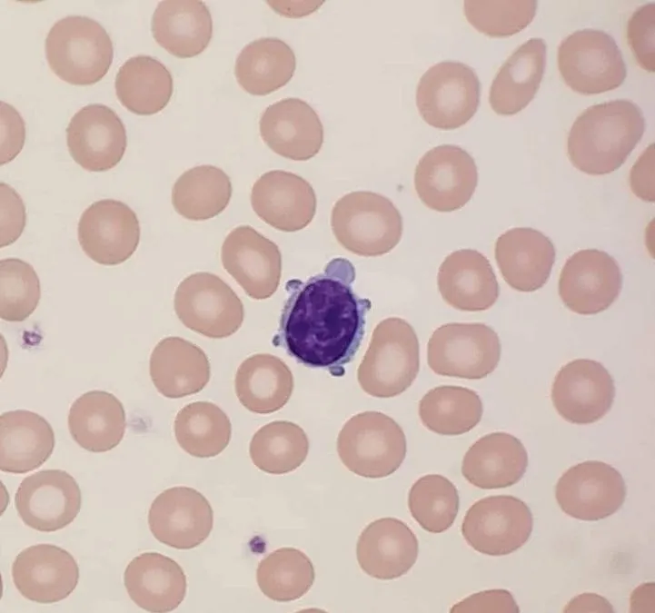
Look at this turtle looking lymphocyte on a peripheral blood smear, looked funny.
If you made it this far reading, thank you for your time.