Krukenberg Tumors are malignancies that involve the ovaries from cancers coming from the gastrointestinal system, the most common organ source would be the stomach. Here's a gross look at the ovaries taken out from the patient on their late 40s.
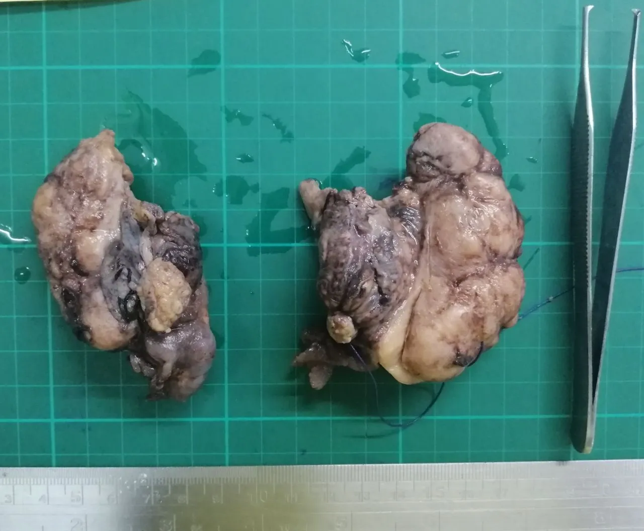
Signs of the tumor being metastatic grossly:
- The ovarian surface looks like it has a lot of implants
- Bilateral involvement
- Multiple organs suspected involved.
On this case, the manifestations were confined to the ovaries. These tumors often spell poor prognosis because their manifestations are discovered when they have already spread, on this case, its the patient's ovaries. I wasn't able to take photo the cut surface but Krukenberg tumors are fairly common enough. Like most stuff I post, anyone can just Google the medical related content. My posts are just written for show and tell.
Taken at scanner, low power view, and high power view respectively:
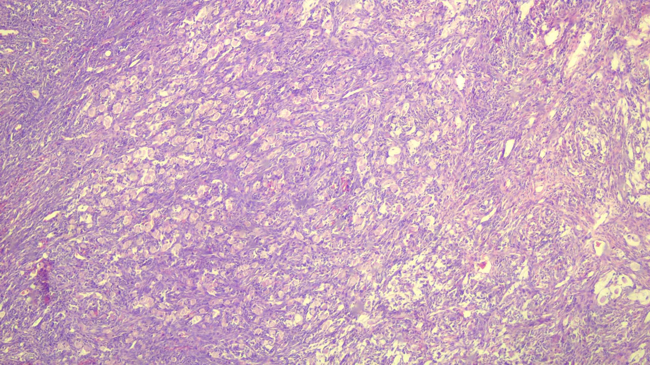
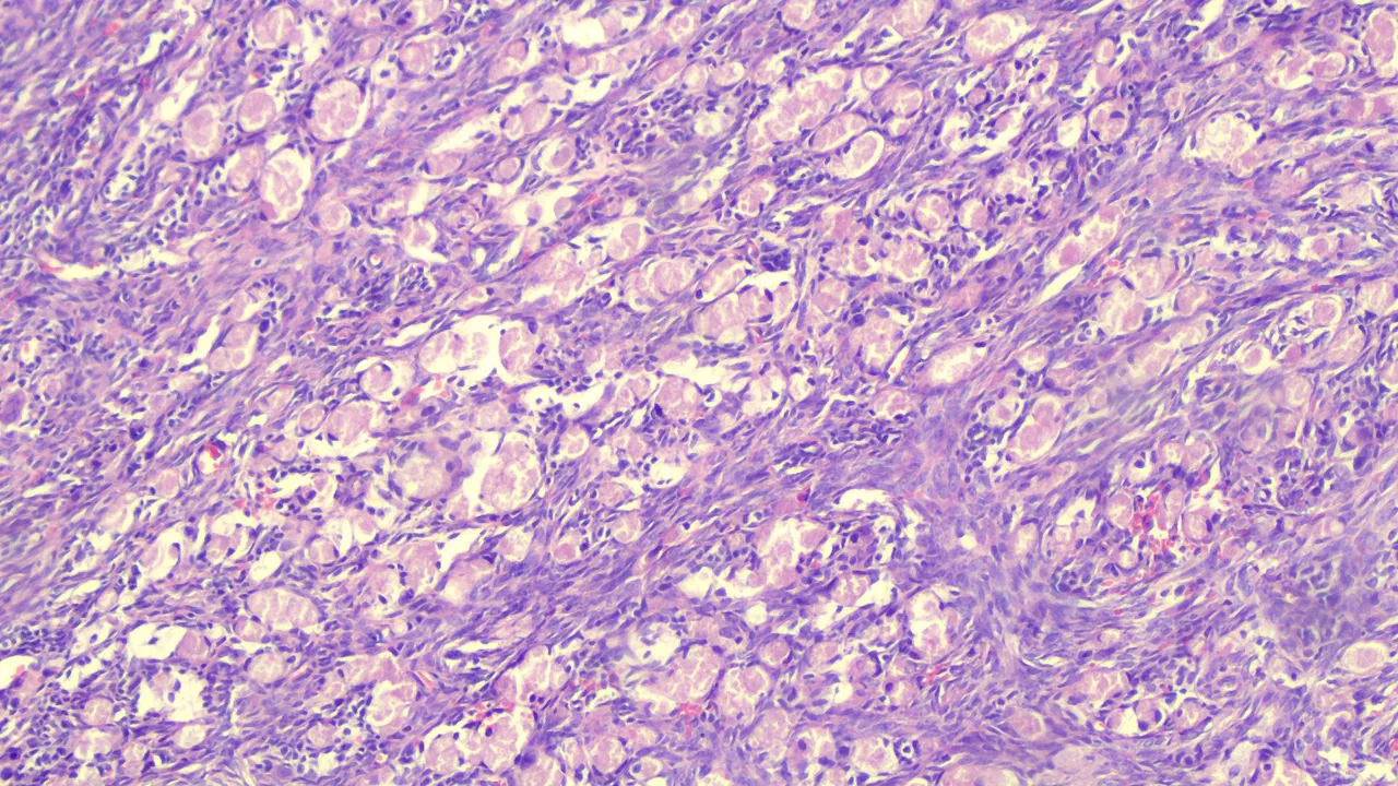
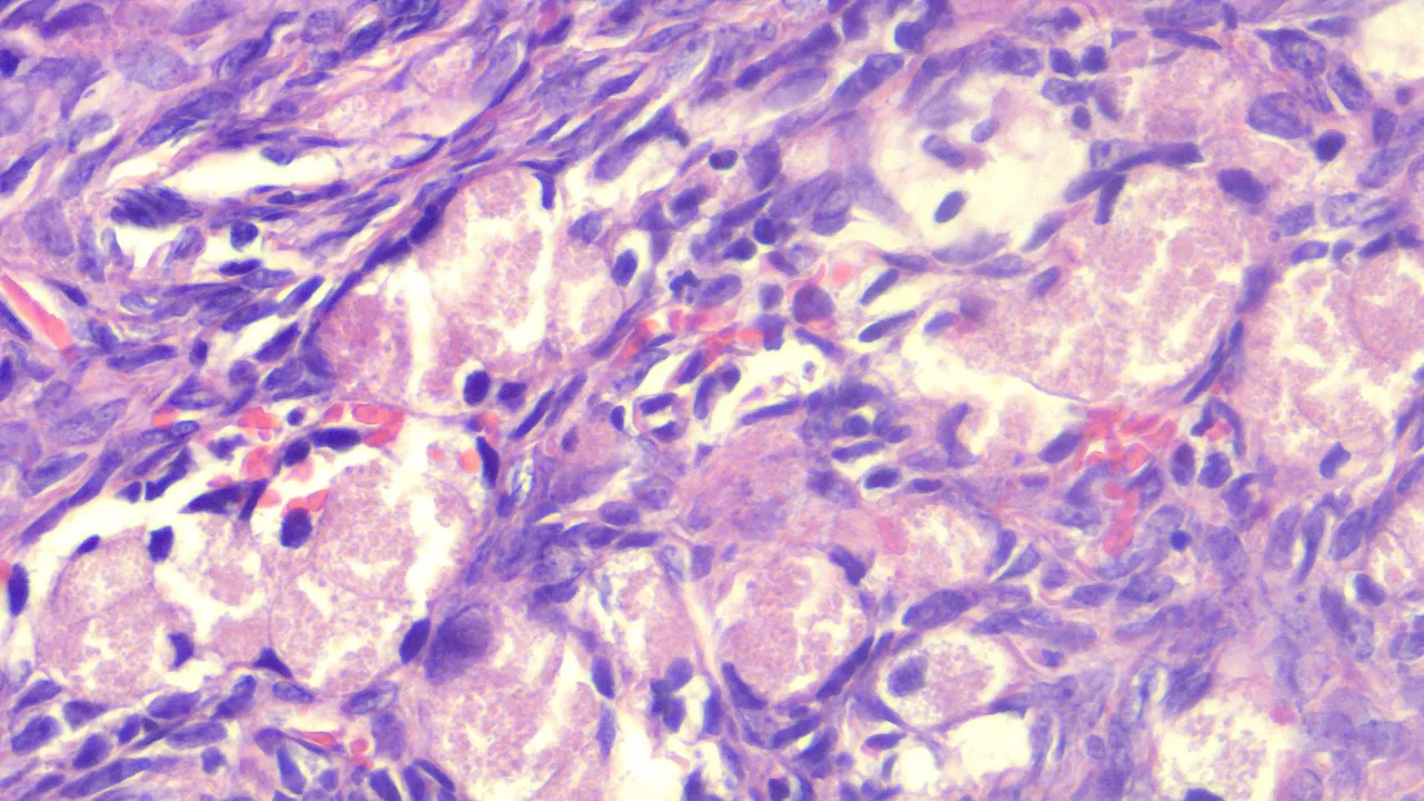
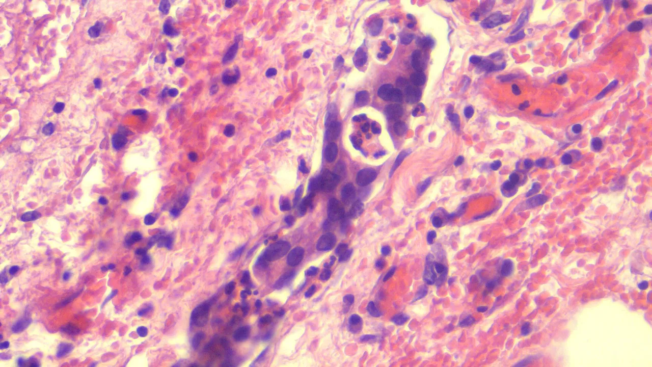
The large granular pink (eosinophilic) to clear cells look like signet ring with the hyperchromatic nucleus displaced at the periphery infiltrating the ovarian tissue arranged in sheets.
Metastatic tumor cells don't necessarily appear like their source tumors but when they accumulate in another site, they may form structures similar to the primary tumor.
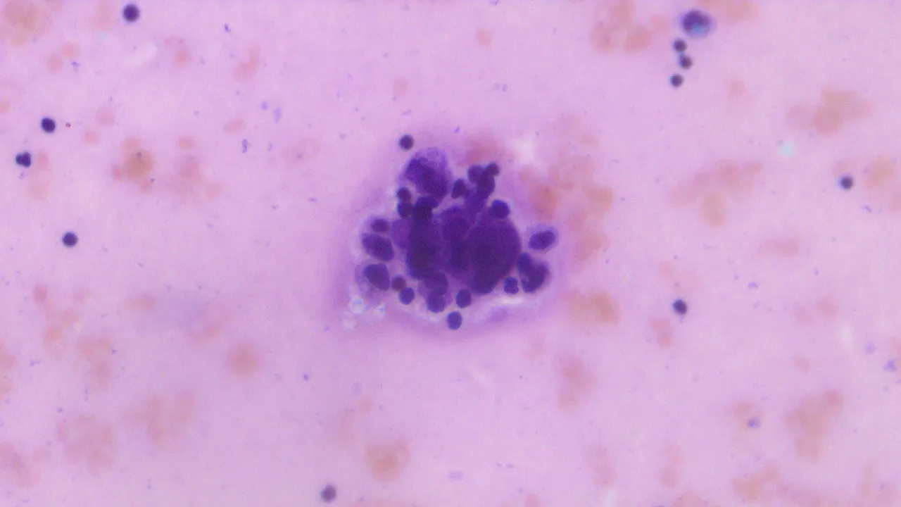
Here's a cluster of tumor cells from the patient's peritoneal fluid:
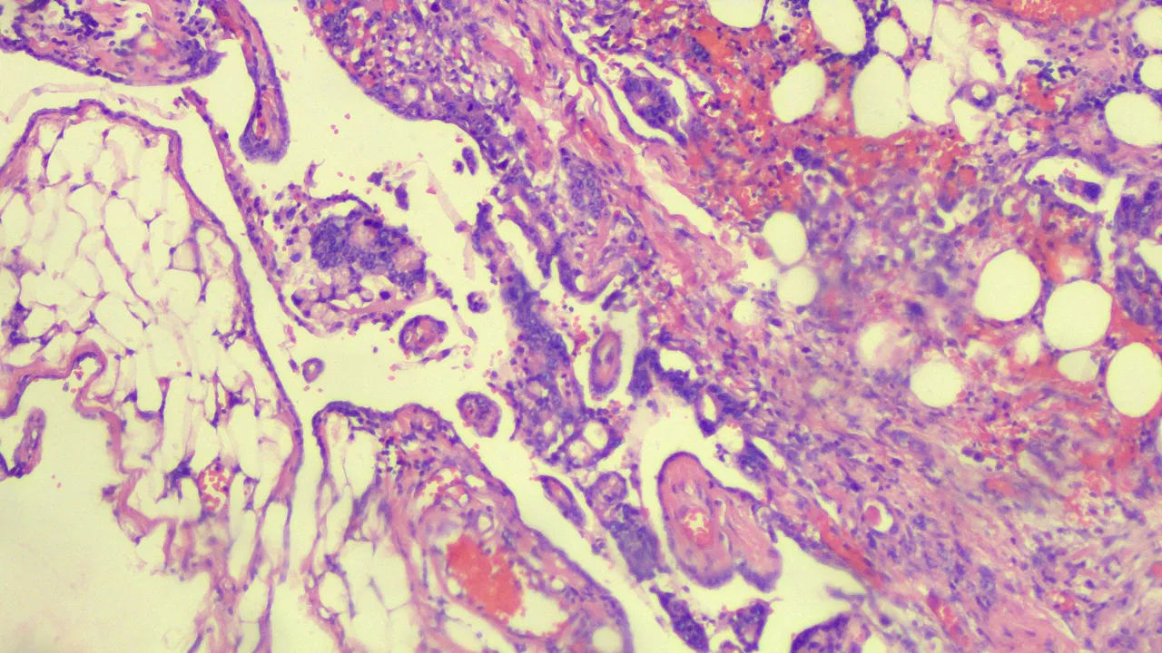
Tumors cells invading the omentum in the middle taken at low power view:
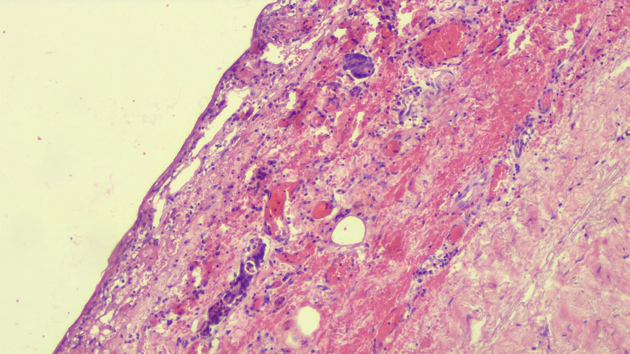
Tumor involving the peritoneum at low power view and high power view:
I wasn't able to take a photo of the immunohistochemical studies done on this patient but it turned out positive for CK7,CK20, CA19-9 and negative for Mammoglobin. This means other than confirming the metastatic cells came from the Gastrointestinal Organs, it's also hinting that stomach or pancreas may be the primary source. The negative mammoglobin was just trying to rule out the malignancy came from the breast.
The patient died before they could perform an endoscopic exam to check the duodenal area. No autopsy was requested to confirm the source. This has been a prevailing problem with the culture here as folks tend to see autopsies as one way of not letting the dead be laid to rest and prolonging the agony.
It's the first time I encountered this case and it wasn't a hard diagnosis to come up with. The next differential diagnosis includes the benign Signet Ring Stromal Tumors and related Sertoli-Leydig Tumors. Seeing the other specimens positive for tumor was more than enough to clinch the diagnosis.
If you made it this far reading, thank you for your time.