Showcasing how clear cell carcinomas look across different organs involved. Please note that this is a phenotypic expression where neoplastic cells have a characteristic hyperchromatic central to eccentric nuclei and abundant clear cytoplasm. These can have a different aberration at a genetic and molecular level but the end expression makes them look like clear cells. I only have three organs for this post from Ovaries, Kidney and Brain specimens.
I'm not going to go into details about how these came to be or prognosis. The post is simply sharing how the same characteristic of neoplastic cells are visibly the almost the same and yet found on different organ locations bearing different clinical implications.
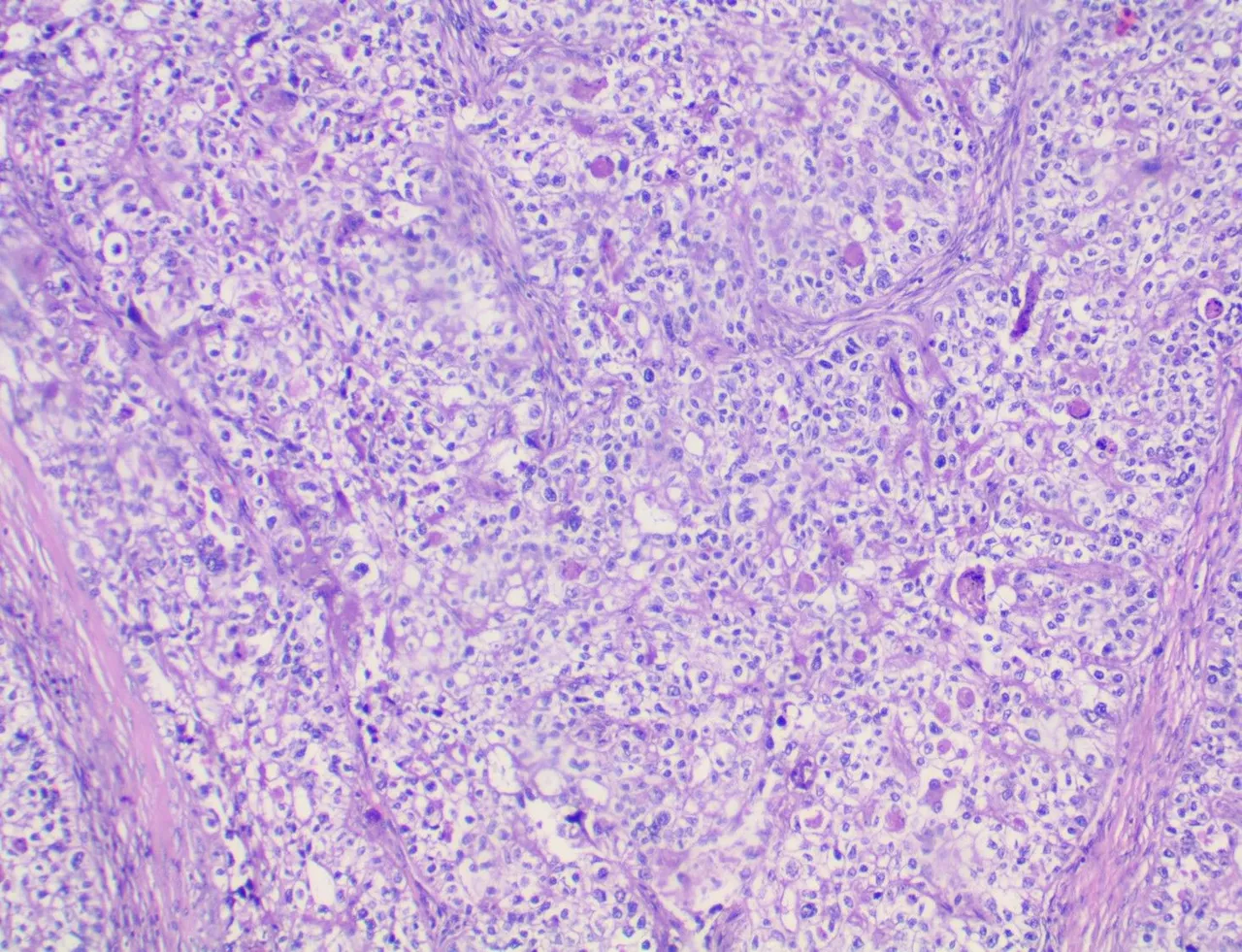
Clear Cell Carcinoma of the Ovary
Taken at Low Power View (100x):
Taken at High Power View (400x)
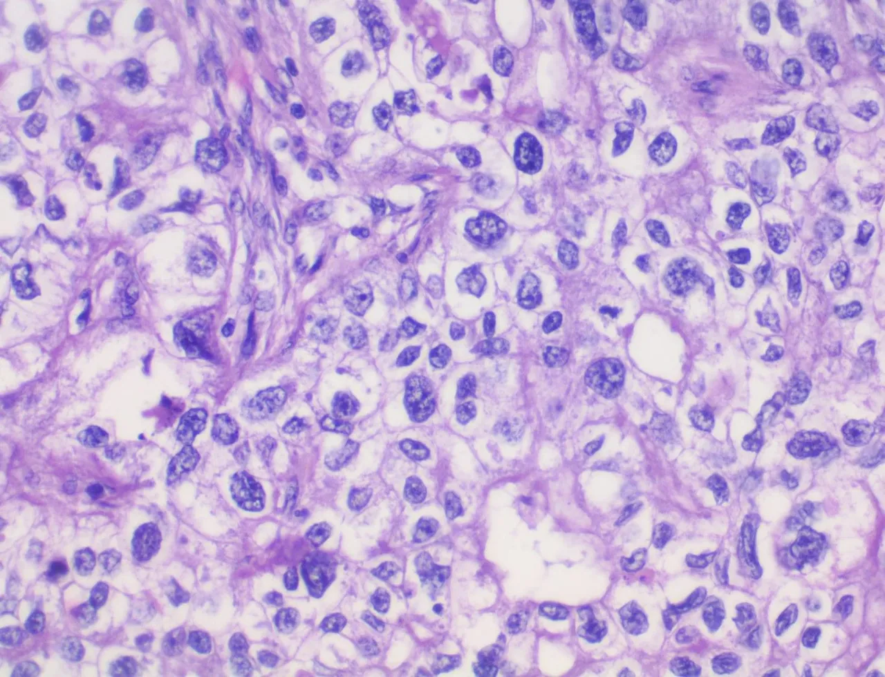
I've never encountered this case before and the pictures came from a colleague's case which I borrowed for study.
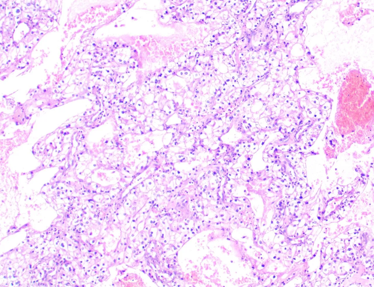
Renal Clear Cell Carcinoma
Taken at Low Power Views (100x)
Taken an High Power View (400x)
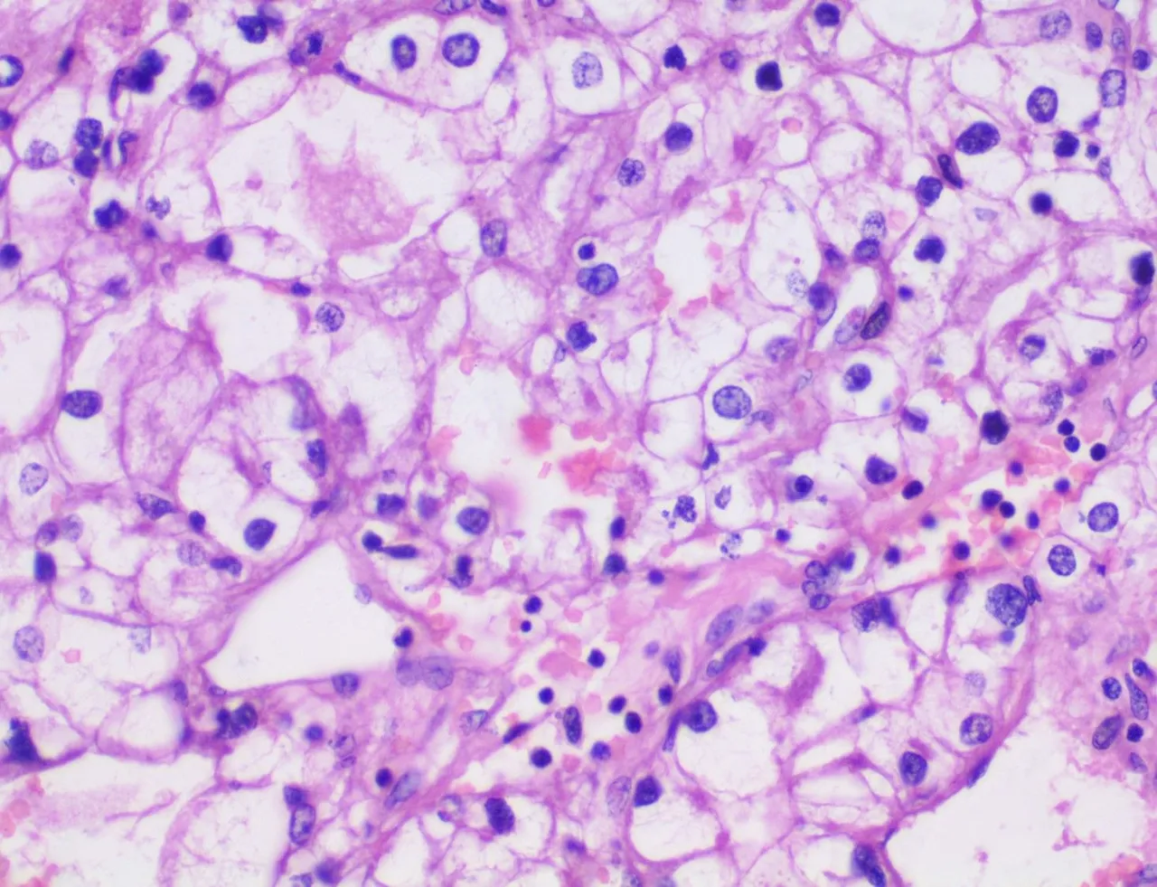
I don't get to cut a lot of radical nephrectomy specimens so I've only seen two cases with renal clear cell carcinoma twice in my training.
A throwback on the Hemangioblastoma case with clear cells.
Taken at Low Power View (100x)

Taken at High Power View (400x)
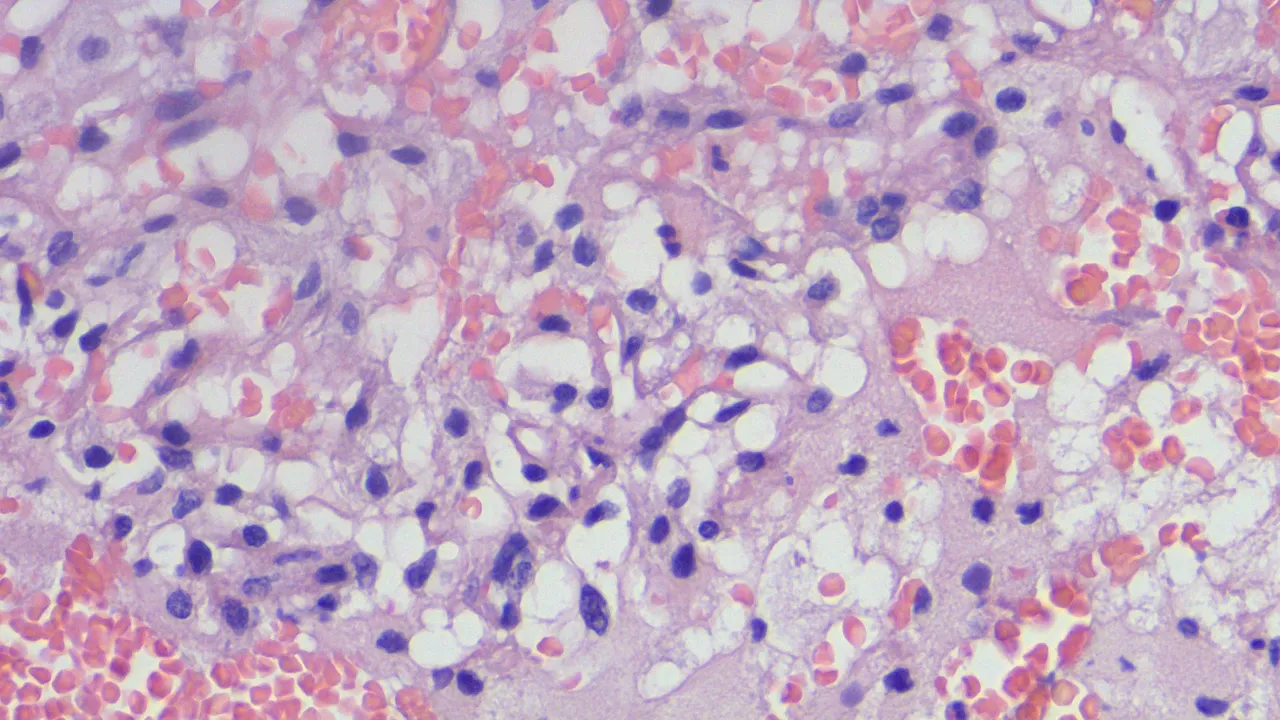
I mentioned before that committing to Hemangioblastoma requires ruling out possible metastatic renal clear cell carcinoma metastasis because you can see the Renal Clear Cell Carcinoma above as an example of how morphology isn't enough sometimes.
Now imagine receiving a slide looking at these features and then no one tells you the origin of the specimen?
If you made it this far reading, thank you for your time.